Rabbit Anti Rat IL-1b polyclonal, antigen affinity
Category: Antigen Affinity Purified$150.00 – $190.00
Description
Accession
Q63264
Applications
ELISA:This antibody can be used at 2 μg/mL jointly with biotinylated Rat IL-1b antibody to detect Rat IL-1b. The detection limit for recombinant Rat IL-1b is approximately 0.2 ng/well.
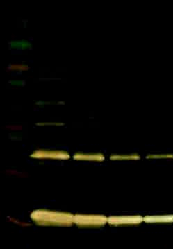
Western Blot:This antibody can be used at 0.2 μg/mL with the appropriate secondary reagents to detect Rat IL-1b. The detection limit for recombinant Rat IL-1b is approximately 0.2 ng/lane.
Source
This antibody was produced in rabbits immunized with recombinant Rat IL-1b.
Species reactivity
Rat
Purification
The specific antibody was purified from Rabbit sera by using immobilized recombinant Rat IL-1b affinity chromatography.
Presentation
Lyophilized from PBS, pH 7.2.
Storage
The lyophilized antibody is stable for at least 1 year from date of receipt at -20° C. Upon reconstitution, this antibody can be stored in working aliquots at 2° - 8° C for one month, or at -20° C for 12 months without detectable loss of activity. Avoid repeated freeze/thaw cycles.
Usage
This antibody product is for research purposes only.It may not be used for therapeutics or diagnostic purposes.
Biological Process
Molecular function
Molecular function
Molecular function
Methods
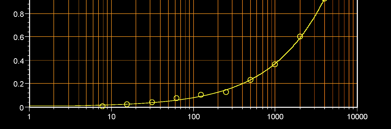
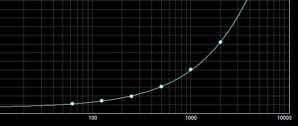
Sandwich ELISA
- 96-well maxisorp plates were coated with the capture antibody overnight, then blocked with 5% FCS in PBS for 1 hour, then washed.
- Then, the protein standard was added, and plates were incubated for 2 hrs at 37°C.
- Plates were washed and incubated with a biotinylated detection antibody for 1 hour at 37°C.
- After washing, plates were incubated with HRP-conjugated streptavidin for 30 min at room temperature and washed again.
- Plates were developed using the tetramethylbenzidine peroxidase substrate system and absorbance was measured at 450-615 nm using an automated plate reader.
Western Blot
- The Recombinant immunogen was migrated by 12% SDS-PAGE and transferred to a 0.2 um PVDF membrane.
- After blocking the membrane in blocking buffer (5% milk powder in 20 mM Tris-HCl pH 7.5, 500 mM NaCl, 0.1% (v/v) Tween 20 , the membrane was incubated with primary antibody ( at 4°C overnight.
- Peroxidase-linked secondary anti-rabbit were used to detect the bound primary antibodies.
- Enhanced chemiluminescence (ECL) reagents were used to visualize