Description
Accession
O35235
Source
Optimized DNA sequence encoding MouseRANK LigandTNF binding domainwas expressed in Escherichia Coli.
Molecular weight
Native Mouse RANK Ligand is a type II transmembrane protein containing an extracellular TNF binding domain, theTNF binding domainhas a calculated molecular mass of approximately20 kDa. Recombinant mouse RANKL is a monomeric protein consisting of amino acid residue subunits. Recombinant RANKLmigrates as an approximately20 kDa protein under non-reducing conditions and reducing conditions in SDS-PAGE.
Purity
>95%, as determined by SDS-PAGE and HPLC
Biological Activity
The ED(50) determined by the dose-dependent stimulation of IL-8 production in Human PBMCs is ≤8 ng/ml.
Protein Sequence
MRRASRDYGK YLRSSEEMGS GPGVPHEGPL HPAPSAPAPA PPPAASRSMF LALLGLGLGQ VVCSIALFLY FRAQMDPNRI SEDSTHCFYR ILRLHENAGL QDSTLESEDT LPDSCRRMKQ AFQGAVQKEL QHIVGPQRFS GAPAMMEGSW LDVAQRGKPE AQPFAHLTIN AASIPSGSHK VTLSSWYHDR GWAKISNMTL SNGKLRVNQD GFYYLYANIC FRHHETSGSV PTDYLQLMVY VVKTSIKIPS SHNLMKGGST KNWSGNSEFH FYSINVGGFF KLRAGEEISI QVSNPSLLDP DQDATYFGAF KVQDID
Endotoxin
Endotoxin content was assayed using a LAL gel clot method. Endotoxin level was found to be less than 0.1 ng/µg(1EU/µg).
Presentation
Recombinant mouse RANK Ligand was lyophilized from a 0.2 μm filtered PBS solution.
Reconstitution
A quick spin of the vial followed by reconstitution in distilled water to a concentration not less than 0.1 mg/mL. This solution can then be diluted into other buffers.
Storage
The lyophilized protein is stable for at least years from date of receipt at -20° C. Upon reconstitution, this cytokine can be stored in working aliquots at2° -8° C for one month, or at -20° C for six months, with a carrier protein without detectable loss of activity. Avoid repeated freeze/thaw cycles.
Usage
This cytokine product is for research purposes only.It may not be used for therapeutics or diagnostic purposes.
Interactor
Interactor
Interactor
O35305
Interactor
P46414
Biological Process
Molecular function
Molecular function
Molecular function
Methods
Osteoblast, osteoclast and macrophage differentiation.
- Bone marrow derived monocytes were seeded in 24-well culture plates and induced to differentiate into macrophages or osteoclasts via the addition of M-CSF or M-CSF + RANKL respectively.
Osteoclastogenesis assays
- Osteoclastogenesis assays were performed as in 5 cells were plated in each well of a 24-well dish with 60 ng/mL RANKL for 3 days.
- After 3 days, 2×10³ MDA-231, shControl, or shEGFR-MDA-231 cells were plated with the bone marrow cells, with 60 ng/mL RANKL and 10 ng/mL MCSF and grown for 3 days before TRAP staining.
- For TRAP staining, cells were washed with 1×PBS, fixed with ice cold methanol for 10 minutes, and stained in fresh TRAP solution for 15 minutes at 37°C.
- TRAP solution was replaced with 1×PBS for cell counting under the microscope.
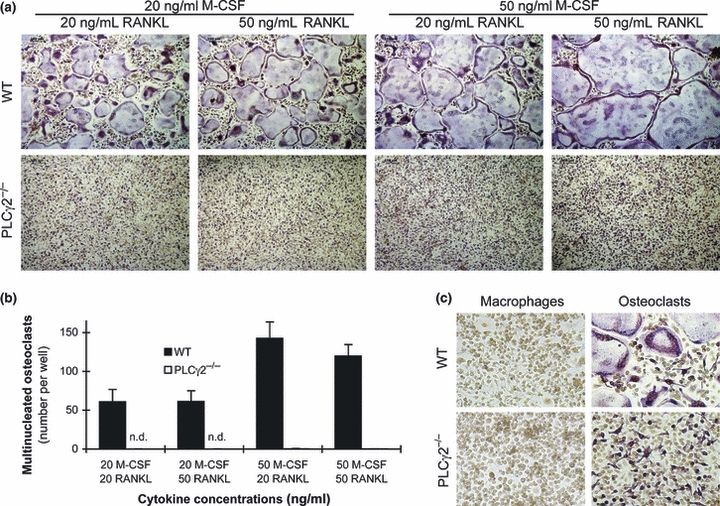
PLCγ2 is required for in vitro osteoclast development.
- (a) Representative TRAP-stained images of wild-type and PLCγ2−/− bone marrow cells cultured in the presence of the indicated concentrations of recombinant murine M-CSF and RANKL for 4 days.
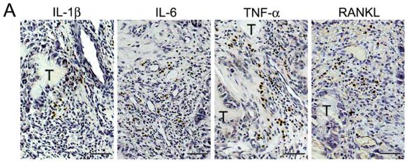
Expression of pro-resorptive cytokines in gouty tophus tissues.
- (c) Immunofluorescent stains for CD3 (green) and RANKL (red) in tophaceous gout tissues.
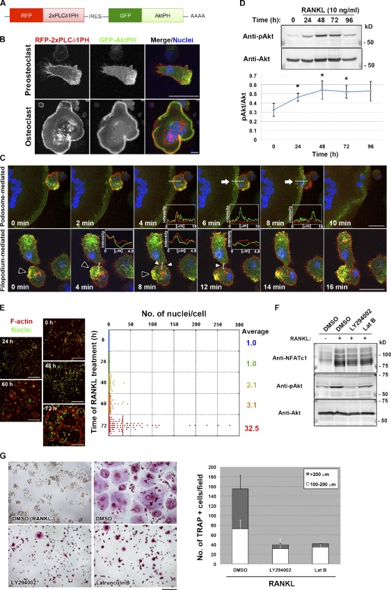
Polarized membrane extensions mediate osteoclast fusion.
- (D, top) Immunoblot analysis of lysates of RAW264.7 macrophages stimulated with RANKL for the indicated times with antibodies to Ser473-phosphorylated (p) or total forms of Akt.
Osteoclast differentiation assay
- Osteoclast differentiation assay was performed as we described.+ T cells or Treg cells (1 × 10⁶/ml) were pre-activated with 2.5 μg/ml anti-CD3 and 1.25 μg/ml anti-CD28 monoclonal mouse antibodies for 12 h as we described,,
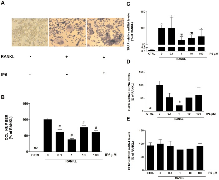
Inositol hexakisphosphate (IP6) directly inhibits osteoclast formation induced by RANKL.
- RAW 264.7 cells cultured for 5 days with no stimulation with RANKL (left).
Inositol hexakisphosphate (IP6) directly inhibits osteoclast formation induced by RANKL.
- RAW 264.7 cells cultured for 5 days with no stimulation with RANKL (left).
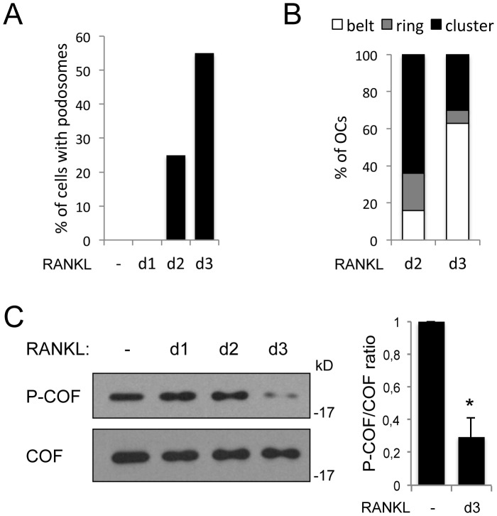
Cofilin is activated when podosome belts are assembled.
- BMMs were cultured for 3 days in the absence or in the presence (d1 to d3) of RANKL.