Mouse Platelet-derived Growth Factor BB Recombinant
Category: Recombinant Mouse Cytokines$70.00 – $4,700.00
Description
Accession
P31240
Source
Optimized DNA sequence encoding mouse PDGF-BB mature chain was expressed in Escherichia Coli.
Molecular weight
MousePDGF-BB, generated by the proteolytic removal of the signal peptide and propeptide. The molecule has a calculated molecular mass of approximately 12 kDa. Recombinant PDGF-BB is a disulfide-linked homodimeric protein consisting of two 109 amino acid residue subunits, and migrates as an approximately 24 kDa protein under non-reducing and reducing conditions in SDS-PAGE.
Purity
>95%, as determined by SDS-PAGE and HPLC.
Biological Activity
The ED(50) was determined by the dose-dependent proliferation of Balb/cT3 cells was found to be in the range of.0-2.0 ng/ml.
Protein Sequence
MNRCWALFLP LCCYLRLVSA EGDPIPEELY EMLSDHSIRS FDDLQRLLHR DSVDEDGAEL DLNMTRAHSG VELESSSRGR RSLGSLAAAE PAVIAECKTR TEVFQISRNL IDRTNANFLV WPPCVEVQRC SGCCNNRNVQ CRASQVQMRP VQVRKIEIVR KKPIFKKATV TLEDHLACKC ETIVTPRPVT RSPGTSREQR AKTPQARVTI RTVRIRRPPK GKHRKFKHTH DKAALKETLG A
Endotoxin
Endotoxin content was assayed using a LAL gel clot method. Endotoxin level was found to be less than 0.1 ng/µg(1EU/µg).
Presentation
Recombinant PDGF-BB was lyophilized from a 0.2 μm filtered solution in.5% glycine,.5% sucrose,.01% Tween80, mM Glutamic acid, pH.5.
Reconstitution
A quick spin of the vial followed by reconstitution in distilled water to a concentration not less than 0.1 mg/mL. This solution can then be diluted into other buffers.
Storage
The lyophilized protein is stable for at least years from date of receipt at -20° C. Upon reconstitution, this cytokine can be stored in working aliquots at2° -8° C for one month, or at -20° C for six months, with a carrier protein without detectable loss of activity. Avoid repeated freeze/thaw cycles.
Usage
This cytokine product is for research purposes only.It may not be used for therapeutics or diagnostic purposes.
Molecular function
Molecular function
Molecular function
Methods
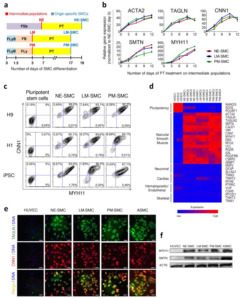
Efficient differentiation of intermediate lineages into vascular SMCs.
- The SMC subtypes, namely the neuroectoderm-derived SMC, lateral mesoderm-derived SMC, paraxial mesoderm-derived SMC are abbreviated as NE-SMC, LM-SMC and PM-SMC respectively.
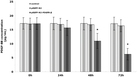
ELISA of PDGF-BB secreted by EPCs in response to PDGFR-β transfection.
- The concentration of PDGF-BB in the supernatant of culture medium was measured using ELISA.
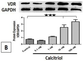
Effect of calcitriol stimulation on VDR mRNA transcript, VDR protein expression, cell proliferation in PCASMCs.
- Quiescent PCASMCs were stimulated with 20 ng/ml PDGF-BB with calcitriol for 24 h, and BrdU incorporation was analyzed .
Growth factor treatment and cell proliferation assay
- Undifferentiated MVSCs and partially differentiated MVSCs (cultured in DMEM with 10% FBS for 3 weeks) were starved in DMEM with 1% FBS for 24 hours followed by the treatment of 10 ng/ml bFGF , 10 ng/ml PDGF-B or 10 ng/ml TGF-β1 for another 24 hours.
- The cell proliferation was quantified by using Click-iT EdU Alexa Fluor 488 HCS Assay kit according to the instruction of the manufacturer.
Primary Culture of Epicardial Cells and Cardiomyocytes
- Primary epicardial cells were cultured in 10% FBS/MEM containing recombinant soluble factors, such as TGFβ1 (10 ng/ml&systems), FGF2 (100 ng/mloche), , , , , PGF-BB (100 ng/ml) or retinoic acid (1 µmol/l), for 3 days.
- The soluble factors were added on the day of heart removal.
- The medium was replaced daily.
- For western blot analysis, cells were treated with TGFβ1 for 2 days, beginning on the day following heart removal, at a final concentration of 1 ng/ml.
In vitro culture of stromal cells
- Stromal cells were derived from E14.5 p190-B−/− and WT fetal livers.
- The cells were cultured in Iscove's Modified Dulbecco's Medium (IMDM) containing 20% fetal bovine serum (FBS, Omega Scientific, Tarzana, CA) and beta mercaptoethanol (2-ME , , ).
- The adherent cells were grown to 70-80% confluence, passaged and sub-cultured in medium containing EGF (10ng/ml ) and PDGF (20ng/ml) .
- The cells were depleted for CD11b using lineage depletion kit as per the manufacturer's protocol.
- All experiments were performed on primary stroma of early passages (p3-p7) and exhibiting similar immunophenotype between the genotypes.
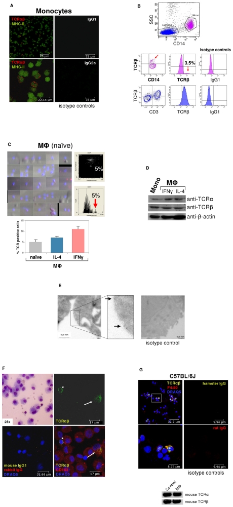
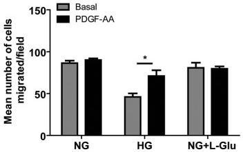
The effects of various glucose conditions on PDGF-mediated migration of retinal AC.
- Transwell assay was performed with basal medium or medium containing PDGF-AA or PDGF-BB in lower compartment.
The effects of various glucose conditions on PDGF-mediated migration of retinal AC.
- Transwell assay was performed with basal medium or medium containing PDGF-AA or PDGF-BB in lower compartment.
In vitro differentiation of Nes-GFP+ cells
-
LC differentiation For LC lineage differentiation, theNes -GFP+ cells were replated in fresh differentiation-inducing medium containing phenol red-free DMEM/F12, 2% FCS, 10 ng/ml PDGF-BB , 1 ng/ml LH , 1 nM thyroid hormone , 70 ng/ml insulin-like growth factor 1 (IGF1), and ITS supplement , and they were incubated for 7 days, as previously described. - Differentiation was subsequently confirmed by RT-PCR and immunostaining for LC lineage markers (antibodies shown in