Mouse Oncostatin M Recombinant
Categories: HematopoietinsRecombinant Mouse Cytokines$70.00 – $4,700.00
Description
Accession
P53347
Source
Optimized DNA sequence encoding Mouse Oncostatin-M mature chain was expressed in Escherichia Coli
Molecular weight
Native Mouse Oncostatin-M isgenerated by the proteolytic removal of the signal peptide and propeptide, this molecule has a calculated molecular mass of approximately kDa. Recombinant mouse Oncostatin-M (OSM) is a monomeric protein consisting of 181 amino acid residue subunits, and migrates as an approximately 20 kDa protein under non-reducingand reducing conditions in SDS-PAGE.
Purity
>95%, as determined by SDS-PAGE and HPLC
Biological Activity
The ED(50) was determined by the dose-dependent stimulation of the proliferation of MouseT3 cells is ≤0.5 ng/ml, corresponding to a specific activity of ≥2 x10⁷ units/mg.
Protein Sequence
MQTRLLRTLL SLTLSLLILS MALANRGCSN SSSQLLSQLQ NQANLTGNTE SLLEPYIRLQ NLNTPDLRAA CTQHSVAFPS EDTLRQLSKP HFLSTVYTTL DRVLYQLDAL RQKFLKTPAF PKLDSARHNI LGIRNNVFCM ARLLNHSLEI PEPTQTDSGA SRSTTTPDVF NTKIGSCGFL WGYHRFMGSV GRVFREWDDG STRSR RQSPL RARRKGTRRI RVRHKGTRRI RVRRKGTRRI WVRRKGSRKI RPSRSTQSPT TRA
Endotoxin
Endotoxin content was assayed using a LAL gel clot method. Endotoxin level was found to be less than 0.1 ng/µg(1EU/µg).
Presentation
Mouse OSM (oncostatin-M) was lyophilized from a 0.2 μm filtered solution in.5% glycine,.5% sucrose,.01% Tween80, mM Glutamic acid, pH.5.
Reconstitution
A quick spin of the vial followed by reconstitution in distilled water to a concentration not less than 0.1 mg/mL. This solution can then be diluted into other buffers
Storage
The lyophilized protein is stable for at least years from date of receipt at -20° C. Upon reconstitution, this cytokine can be stored in working aliquots at2° -8° C for one month, or at -20° C for six months, with a carrier protein without detectable loss of activity. Avoid repeated freeze/thaw cycles.
Usage
This cytokine product is for research purposes only.It may not be used for therapeutics or diagnostic purposes.
Interactor
Interactor
Biological Process
Molecular function
Methods
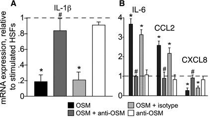
Impact of OSM on gene expression (A, B) and protein secretion (C, D) in stimulated HSFs.
- Human recombinant OSM (10 ng/ml) was added with or without 10 μg/ml of neutralizing anti-OSM, or isotype-matched non-specific antibody.
Isolation and culture of Nestin-positive cells from Nes-GFP transgenic mice
-
The testes were dissected from 7-day-old
Nes -GFP or C57BL/6 mice, and the tunica albuginea was carefully removed from each testis and minced into small pieces. - The interstitial cells were then dissociated from the seminiferous tubules by digestion with 1 mg/ml collagenase type IV in medium'>Dulbecco's medium'>modified medium'>Eagle's medium (DMEM)/F12 at 37 °C for 15 min.
- After adding DMEM containing 5% fetal bovine serum (FBS, Hyclone) to stop the collagenase activity, the samples were centrifuged at 1 500 rpm for 5 min at room temperature; the pellets were washed twice with phosphate-buffered saline (PBS), resuspended in PBS, and filtered through a 45 μm filter, thereby excluding seminiferous tubules from the preparation.
- The suspension was passed through a 20 μm strainer, which resulted in single cells.
- GFP-expressing cells that exhibited fluorescence intensities ∼10-fold greater than the autofluorescence of cells from C57BL/6L mice were enriched…