Mouse Interleukin-5 Recombinant
Categories: HematopoietinsIL-3 familyRecombinant Mouse Cytokines$70.00 – $4,700.00
Description
Accession
P04401
Source
Optimized DNA sequence encoding Mouse Interleukin-5 mature chain was expressed in Insect Cells
Molecular weight
Native Mouse Interleukin-5, generated by the proteolytic removal of the signal peptide and propeptide, the molecule has a calculated molecular mass of approximately 13 kDa. Recombinant mouse IL-5 is a disulfide-linked homodimeric protein consisting of two 113 amino acid residue subunits,and migrates as an approximately 26 kDa protein under non-reducing conditions and as a 13 kDa protein under reducing conditions in SDS-PAGE.
Purity
>95%, as determined by SDS-PAGE and HPLC
Biological Activity
The ED(50) was determined by the dose-dependent proliferation of TF-1 cells was ≤.2 ng/ml, corresponding to a specific activity of ≥ x units/mg.
Protein Sequence
MRRMLLHLSV LTLSCVWATA MEIPMSTVVK ETLTQLSAHR ALLTSNETMR LPVPTHKNHQ LCIGEIFQGL DILKNQTVRG GTVEMLFQNL SLIKKYIDRQ KEKCGEERRR TRQFLDYLQE FLGVMSTEWA MEG
Endotoxin
Endotoxin content was assayed using a LAL gel clot method. Endotoxin level was found to be less than 0.1 ng/µg(1EU/µg).
Presentation
Recombinant Interleukin-5 was lyophilized from a 0.2 μm filtered PBS solution.
Reconstitution
A quick spin of the vial followed by reconstitution in distilled water to a concentration not less than 0.1 mg/mL. This solution can then be diluted into other buffers.
Storage
The lyophilized protein is stable for at least years from date of receipt at -20° C. Upon reconstitution, this cytokine can be stored in working aliquots at2° -8° C for one month, or at -20° C for six months, with a carrier protein without detectable loss of activity. Avoid repeated freeze/thaw cycles.
Usage
This cytokine product is for research purposes only.It may not be used for therapeutics or diagnostic purposes.
Molecular function
Molecular function
Methods
γIMCs are the source of IFN-γ in severe invasive GAS infections.
- (f) Sorted IMCs were cultured with control medium (Med), G-CSF (50 ng ml−1), M-CSF (10 ng ml−1), GM-CSF (10 ng ml−1), or IL-5 (10 ng ml−1) for 2–4 days, and their absolute numbers were counted on the indicated days.
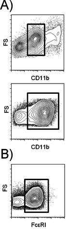
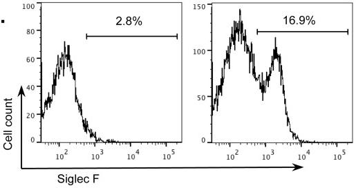
CD200R and CD200RLc expression on mouse leukocytes.
- Cells cultured in IL-5 supplemented media were gated as CD11bSiglec F eosinophils in CD11b-Siglec F plot (bottom left).
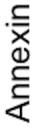
Effects of DPI on Siglec-F-induced apoptosis of eosinophils.
- Wild-type mouse bone marrow-derived eosinophils (n = 4) were incubated with or without DPI (20 µM) for 5 min before adding either a control mAb or Siglec-F mAb and then cells were cultured in IL-5 for an additional 18 hr before determining Annexin positivity.
Expansion of bmEos ex vivo.
- Bone marrow cells from BALB/c mice were cultured for 14 days, see Methods and samples taken from day 4 to day 14 in cultures grown in the presence of IL-5.
Generation of bone marrow-derived eosinophils
- The cells were cultured at 37°C in the presence of 5% CO2.
- On day 4, the cells were removed from the flasks by pipetting, centrifuged, and resuspended in an equivalent volume of fresh medium'>BMDE medium containing 10 ng/ml IL-5 .
- The cells were then transferred to new flasks and cultured at 37°C in the presence of 5% CO2.
- On day 8, the cells were again removed from the flasks by pipetting, centrifuged, and resuspended in an equivalent volume of fresh medium'>BMDE medium containing 10 ng/ml IL-5 .
- The cells were then transferred to new flasks and cultured at 37°C in the presence of 5% CO2.
- On days 10, 12, 14, and 16, the cells were removed from the flasks by pipetting, centrifuged, counted, adjusted to a concentration of 106 cells/ml in fresh medium'>BMDE medium containing 10 ng/ml IL-5, and cultured at 37°C in…