Mouse Interleukin-12 p40 Recombinant
Categories: HematopoietinsRecombinant Mouse Cytokines$70.00 – $160.00
Description
Accession
P43432
Source
Optimized DNA sequence encoding mouse Interleukin-12 mature chain was over-expressed in CHo cells.
Molecular weight
Recombinant mouse IL-12 is a heterodimer protein of 35kDa +40kDa subunits and consisting of 506 amino acid residue subunits. IL-12 migrates as an approximately 80kDa protein under non-reducing conditions and as 35kDa and 40kDa bands under reducing conditions in SDS-PAGE.
Purity
>95%, as determined by SDS-PAGE and HPLC
Biological Activity
The ED(50) determined by the dose-dependent stimulation of IFN-gamma production by murine splenocytes co-stimulated with IL-18 is <.1 ng/ml, corresponding to a specific activity of > x units/mg.
Protein Sequence
MCPQKLTISW FAIVLLVSPL MAMWELEKDV YVVEVDWTPD APGETVNLTC DTPEEDDITW TSDQRHGVIG SGKTLTITVK EFLDAGQYTC HKGGETLSHS HLLLHKKENG IWSTEILKNF KNKTFLKCEA PNYSGRFTCS WLVQRNMDLK FNIKSSSSSP DSRAVTCGMA SLSAEKVTLD QRDYEKYSVS CQEDVTCPTA EETLPIELAL EARQQNKYEN YSTSFFIRDI IKPDPPKNLQ MKPLKNSQVE VSWEYPDSWS TPHSYFSLKF FVRIQRKKEK MKETEEGCNQ KGAFLVEKTS TEVQCKGGNV CVQAQDRYYN SSCSKWACVP CRVRS
Endotoxin
Endotoxin content was assayed using a LAL gel clot method. Endotoxin level was found to be less than 0.1 ng/µg(1EU/µg).
Presentation
Recombinant Interleukin-12 was lyophilized from a 0.2 μm filtered PBS solution.
Reconstitution
A quick spin of the vial followed by reconstitution in distilled water to a concentration not less than 0.1 mg/mL. This solution can then be diluted into other buffers.
Storage
The lyophilized protein is stable for at least years from date of receipt at -20° C. Upon reconstitution, this cytokine can be stored in working aliquots at2° -8° C for one month, or at -20° C for six months, with a carrier protein without detectable loss of activity. Avoid repeated freeze/thaw cycles.
Usage
This cytokine product is for research purposes only.It may not be used for therapeutics or diagnostic purposes.
Interactor
Interactor
Q99665
Molecular function
Methods
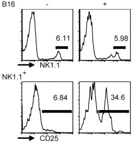
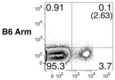
Exhausted CD8 T cells do not upregulate CD25 expression following IL-12, IL-18, and/or IL-21 exposure.
- Representative flow cytometry plots show the expression of CD25 by gated CD8 T cells following acute (B6 Arm), protracted (B6 cl13), and chronic (CD4-/- cl13) LCMV infections in response to a 5.5 hr incubation with or without IL-12, IL-18, and IL-21 in the absence of BFA.
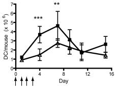
Increased splenic and hepatic DC numbers following systemic IL-12 treatment.
- BALB/c mice were treated with 1 µg/mouse IL-12 (solid square) or vehicle control (VC; open triangle) for 4 consecutive days from day 0, as indicated with arrows, then leukocyte populations analyzed at various days post treatment.
Mouse T Cell Primary Cultures
- Peripheral lymph node cells were pooled from axillary, inguinal, brachial, and cervical lymph nodes (excluding mesenteric (mes)LN).
- Where explicitly stated in the figure legends mesLN were collected.
- After lymphoid organ disaggregation, cells were washed and resuspended in complete media (RPMI-1640, 5%FCS, 2 mM L-gln, 100 U/ml penicillin/streptomycin, and 2.5×10⁻⁵ M 2β-ME .
- Anti-CD3 (145-2C11, BD) at 0-1 µg/ml and anti-CD28 (37.51, BD) at 2 µg/ml mAb were coated on plates (1h, 37°C, in PBS) and washed twice.
- In transient TCR stimulations, cells were moved from Ab-coated to fresh wells on d2.
- aAPC were described previously
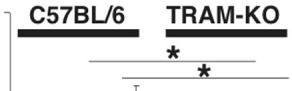
The IFN-γ production from IL-18 and/or IL-12 stimulated Th1 cells from TRAM-deficient mice and MyD88 deficient mice.
- The black bars show the production levels from IL-18 and IL-12 co-stimulated Th1 cells, grey bars show those from IL-18 solely stimulated Th1 cells, and the white bars show those from no secondary stimulated Th1 cells.
In vitro proliferation
- Naive OT-1 T cells were purified using a CD8 isolation kit with supplemental anti-CD44, and labeled with 5 μM CFSE .
- T cell and celltype'>NK cell depleted splenocytes were purified by a similar procedure as OT-1 T cells with an antibody mixture containing biotin-αTCRβ (H57-597), biotin-αTCRγδ (eBioGL3) and biotin-αcelltype'>NK1.1 (pk136) and used as APCs.
- APCs were pulsed with 0.2–5ng/ml peptide (SIINFEKL and all variant peptides were from Genemed Synthesis) and 1µg/ml LPS at 37°C for 1 hour followed by extensive washing.
- OT-1 T cells and APCs were mixed and cultured in the presence of 2.5ng/ml hTGF-β1 (&systems).
- As indicated, 1000U/ml IFN-αA (PBL InterferonSource), 20ng/ml IFN-γ and 20ng/ml IL-12 were added to the culture.
- 50U/ml IL-2 (eBioscience) was added one day later.
- 10µg/ml αTGF-β neutralizing antibody (clone#1D11, R&D systems) was added as indicated in the absence exogenous hTGF-β1.
- To calculate the T cell number during…
T-cell polarization.
- Splenic cells (2 × 10⁶/mL) from age 4 to 6 weeks naïve NOD.BDC2.5.FoxP3GFP.DTR mice were stimulated with p79 peptide (0.5 μmol/L) under Th1 or Th17 conditions for 4 days.
- For polarization into Th1 cells, the stimulation was carried out in the presence of recombinant (r)IL-12 (10 ng/mL) and anti–IL-4 (10 μg/mL [11B11]).
- For Th17 polarization, the culture was supplemented with recombinant transforming growth factor-β (3 ng/mL), rIL-6 (20 ng/mL), anti–IFN-γ (10 μg/mL [4–6A4]), and anti–IL-4 (10 μg/mL [11B11]) antibodies and rIL-23 (20 ng/mL& ) (only for the last 2 days).
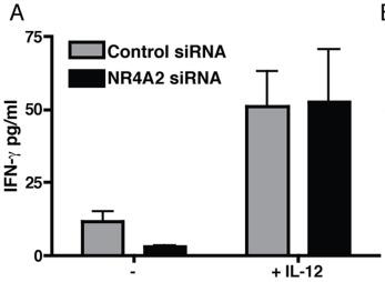
NR4A2 knockdown prevents IL-17 secretion but not RORγt upregulation.
- A: IFN-γ production by cells activated in the presence or absence of 10 ng/ml IL-12 after 96 hours of culture.
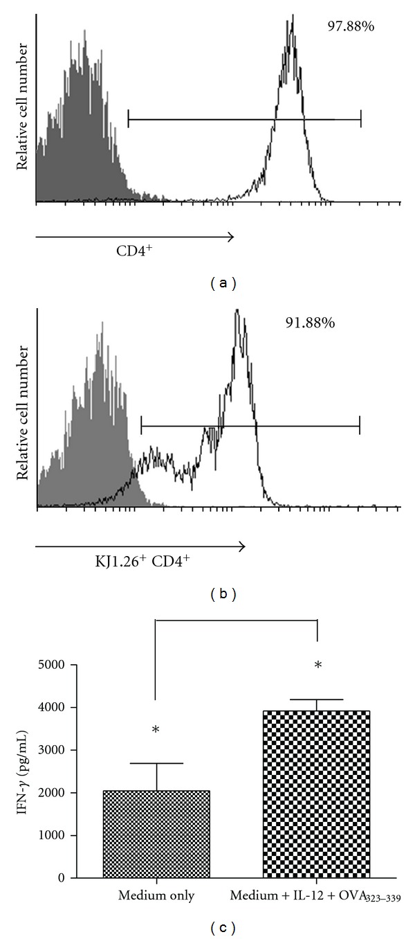
KJ1.26+ CD4+T cell purification and Th1 polarization of DO11.10 cells.
- Th1 polarization, then, was induced in purified cells, (c) IFN-γ levels were measured in the supernatant of cocultures of DO11.10 CD4+ T cells, and macrophages, IL-12, and OVA323-339 peptide were used.
Functional assays with T cells of healthy donors
- celltype'>T cell activation experiments using the system of celltype'>T cell stimulator cells were done as previously described 4/well) were co-cultured with 1×10⁵ celltype'>T cells, and following 48 h of stimulation, cell culture supernatant was harvested for cytokine measurement and methyl-3[H]-thymidine was added to the cultures.
- Expression of membrane-bound anti-CD3 antibody fragments on the T cell stimulator cells was detected via DyLight-649-conjugated goat antibodies to mouse IgG-H+L specific .
- Isolation of human T cells and monocytes and generation of immature and mature monocyte-derived DC was done as previously described 5 T cells/well were added and cultured for 48 h in the presence of soluble TIM-3 antibodies or isotype control antibodies (bothfinal concentration: 5 µg/ml) as previously described 5/well) were co-cultured with allogeneic immature or mature human DC at the indicated cell numbers for 5…
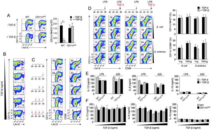
TGF-β suppresses DC production but has no effect on DC activation.
- Bar graphs summarize the average production of IL-12, IL-6, and IL-10 from CD11cdnR (n = 4) and wild-type (n = 4) cultures.