Mouse Interferon-gamma Recombinant
Category: Recombinant Mouse Cytokines$70.00 – $1,000.00
Description
Accession
P01580
Source
Optimized DNA sequence encoding Mouse Interferon gamma mature chain was expressed in Escherichia Coli.
Molecular weight
Native Mouse IFN-g, generated by the proteolytic removal of the signal peptide and propeptide, the molecule has a calculated molecular mass of approximately 16kDa. Recombinant Interferon gamma is a monomer protein consisting of 134 amino acid residue subunits, and migrates as an approximately 16kDa protein under non-reducing and reducing conditions in SDS-PAGE.
Purity
>95%, as determined by SDS-PAGE and HPLC
Biological Activity
The ED(50) was determined by the cytopathic inhibition assay with murine L929 cells challenged with EMC virus was found to be.1 ng/ml
Protein Sequence
MNATHCILAL QLFLMAVSGC YCHGTVIESL ESLNNYFNSS GIDVEEKSLF LDIWRNWQKD GDMKILQSQI ISFYLRLFEV LKDNQAISNN ISVIESHLIT TFFSNSKAKK DAFMSIAKFE VNNPQVQRQA FNELIRVVHQ LLPESSLRKR KRSRC
Endotoxin
Endotoxin content was assayed using a LAL gel clot method. Endotoxin level was found to be less than 0.1 ng/µg(1EU/µg).
Presentation
Recombinant mouse IFN-g was lyophilized from a 0.2 μm filtered PBS solution.
Reconstitution
A quick spin of the vial followed by reconstitution in distilled water to a concentration not less than 0.1 mg/mL. This solution can then be diluted into other buffers.
Storage
The lyophilized protein is stable for at least years from date of receipt at -20° C. Upon reconstitution, this cytokine can be stored in working aliquots at2° -8° C for one month, or at -20° C for six months, with a carrier protein without detectable loss of activity. Avoid repeated freeze/thaw cycles.
Usage
This cytokine product is for research purposes only.It may not be used for therapeutics or diagnostic purposes.
Biological Process
Biological Process
Molecular function
Methods
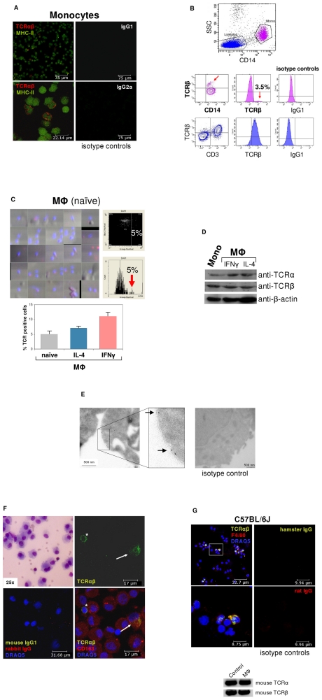
ΤCRαβ expression by subpopulations of human and murine monocytes/macrophages.
- (Bottom) LSC of naïve and IL-4 (10 ng/ml) or IFNγ (1000 U/ml) stimulated monocyte-derived macrophages, respectively, cultured for 6 days.
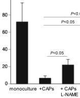
Cardiac adherent proliferating cells reduce Coxsackievirus B3 viral progeny release in a nitric oxide- and interleukin-10-dependent manner and require interferon-γ priming.
- HeLa cells were incubated for 30 minutes with 1 ml of diluted supernatant of CVB3-infected HL-1 cells or HL-1 cells co-cultured with CAPs, in the presence or absence of L-NAME, anti-human IL-10 antibody, or anti-murine IFN-γ antibody, or with diluted LV homogenates from CVB3 or CVB3+CAPs mice.
Cytokine measurements
- The cytokine concentration in the BAL fluid was quantified by ELISA kits specific for IL-5 and IL-10 and for IFN-γ (INC.).
- The values are expressed as picograms per milliliter deduced from standards, run in parallel with the recombinant cytokines.
- The limit of detection values were 10 pg/mL for IL-5 and IL-10 and 16 pg/mL for IFN-γ.
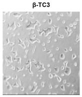
Selection of a β-TC3 cell population resistant to cytokine-mediated cytotoxicity.
- Cytotoxicity assay in β-TC3 (left) and β-TC3R (right) after cytokine treatment (100 IU/ml IL-1β, 100 IU/ml IFN-γ or their combination, for 72 hours).
Cytokine Assays
- ELISAs were also performed to assess levels of interleukin (IL)-2, IL-4, IL-10, and interferon (IFN)-γ in the supernatant of the MLCs on day 4.
- The capture monoclonal antibody (mAb) (JES5-2A5), detection mAb (JES5-16E3), and recombinant standard for IL-10 were from .
- The capture and detection mAbs for IL-2 (JES6-1A12 and JES6-5H4, respectively), IL-4 (BVD-1D11 and BVD-24G2), and IFN-γ (R4-6A2 and XMG1.2) were from .
- Recombinant standards for IL-2, IL-4, and IFN-γ were from .
Cell culture
- Phoenix cells were cultured and maintained in Dulbecco's Modified Eagle's Medium (DMEM supplemented with glutamine, 4.5 g/l glucose), 10% (v/v) fetal bovine serum (FBS) and 1% (v/v) penicillin/streptomycin (PAA) at 37 °C and 5% CO2.
MuSK andMuSK myoblasts isolated from embryos carrying a temperature-sensitive SV40 T antigen were propagated at 33 °C and 5% CO2 on 0.2% gelatin-coated dishes containing growth medium: DMEM containing glutamine, 4.5 g/l glucose enriched with 10% (v/v) FBS, 10% (v/v) horse serum (HS), 0.25% chick embryo extract (CEE), 20 U/ml recombinant murine interferon-γ (IFN-γ) and 1% (v/v) penicillin/streptomycin (MuSK muscle cells was performed according to Herbst and Burden (MuSK myoblasts in the presence of 2 μg/ml polybrene .- After 2 h the virus-containing medium was replaced with fresh growth media.
- 24 h post-infection, muscle cells were split and maintained in growth medium with puromycin (2 ng/ml).
- Clones of cells were…
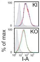
MHC-II surface expression on KI and KO antigen presenting cells is similar to WT
- Thioglycolate elicited peritoneal macrophages were stimulated in vitro 24 h with 500 U/ml IFN-γ.
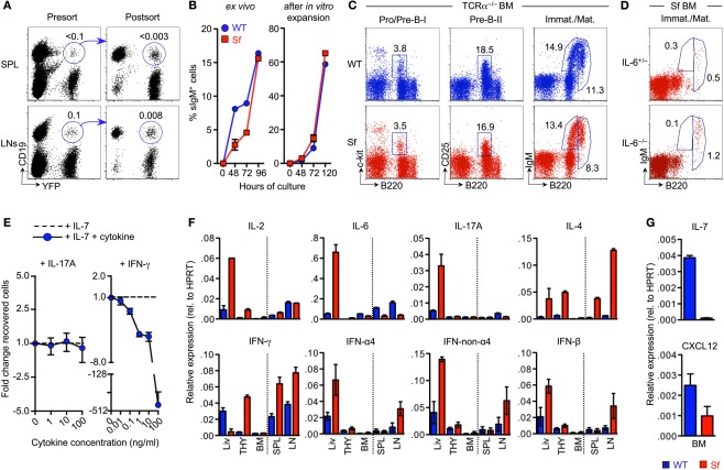
Mechanisms of B cell developmental defects in adolescent Sf mice.
- FACS-purified populations of BM-derived B220+c-kit+ Pro/Pre-B-I cells from WT mice were cultured with IL-7, either alone or in the presence of titrating amounts of IL-17A or IFN-γ, as indicated.
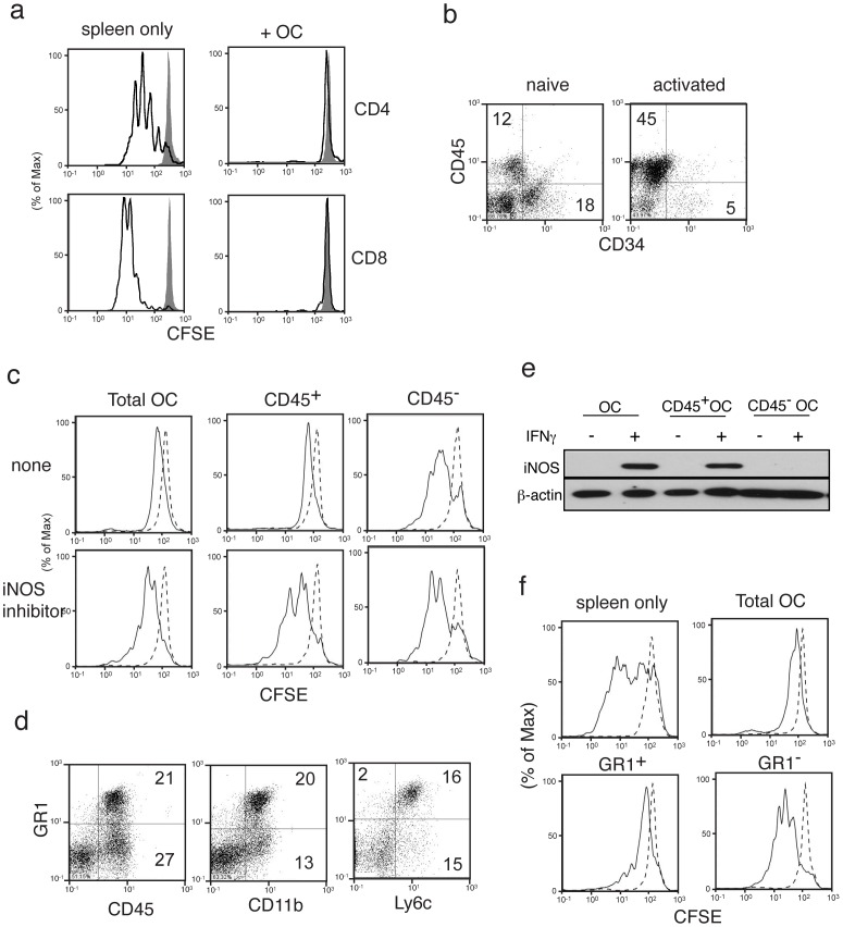
Effect of omentum cells on ex vivo T cell proliferation.
- (e) iNOS expression was determined in total omentum cells, CD45+ omentum cells, or CD45− omentum cells upon IFNγ stimulation for 24 hrs by western blotting.