Mouse GM-CSF Recombinant

Mouse GM-CSF Recombinant

$70.00$2,900.00


accession P01587


Source Optimized DNA sequence encodingMouse Granulocyte Macrophage-Colony Stimulating Factor mature chain was expressed in Escherichia Coli.
Molecular weight Native Mouse Granulocyte Macrophage-Colony Stimulating Factor isgenerated by the proteolytic removal of the signal peptide and propeptide, this molecule has a calculatedmass of kDa. Recombinant mouse GM-CSF is a monomer protein, consisting of amino acidsandmigrates as an approximately14 kDa protein under reducing conditions.
Purity >95%, as determined by SDS-PAGE and HPLC
Biological Activity The ED50 as determined by the dose-dependent stimulation of the proliferation of murine FDC-P1 cells is ≤.1ng/ml, corresponding to a specific activity of ≥3 x units/mg.
Protein Sequence MWLQNLLFLG IVVYSLSAPT RSPITVTRPW KHVEAIKEAL NLLDDMPVTL NEEVEVVSNE FSFKKLTCVQ TRLKIFEQGL RGNFTKLKGA LNMTASYYQT YCPPTPETDC ETQVTTYADF IDSLKTFLTD IPFECKKPGQ K
Endotoxin Endotoxin content was assayed using a LAL gel clot method. Endotoxin level was found to be less than.1 ng/µg(1EU/µg).
Presentation Recombinant mouse GM-CSF was lyophilized from.2 μm filtered PBS solution, pH7.0.
Reconstitution A quick spin of the vial followed by reconstitution in distilled water to a concentration not less than.1 mg/mL. This solution can then be diluted into other buffers.
Storage The lyophilized protein is stable for at least years from date of receipt at -20° C. Upon reconstitution, this cytokine can be stored in working aliquots at° -° C for one month, or at -20° C for six months, with a carrier protein without detectable loss of activity. Avoid repeated freeze/thaw cycles.
Usage This cytokine product is for research purposes only.It may not be used for therapeutics or diagnostic purposes.


Molecular function Cytokine
Molecular function Growth-factor

Methods

In-vitro NK-DC co-culture

  • Whole blood samples were obtained from healthy laboratory donors for the isolation of purified and enriched NK and DC cell populations from PBMC using magnetic cell separation.
  • The development of an NK-DC co-culture system required two independent steps.
  • Firstly, DC were prepared using a pre-optimised magnetic cell sorting kit for the extraction of CD14+ monocytes from PBMC.
  • Following isolation, CD14+ monocytes were treated with 100 ng/ml of GM-CSF and 1000 U/ml of IL-4 in RPMI containing 5% autologous serum and maintained in a 5% CO2 cell culture incubator at 37°C for 5 days.
  • This altered their phenotype to that of immature dendritic cells (iDC) with a population of >90% CD14CD1a+ iDC cells, comparable with the published literature +CD16+ NK cell.
  • Purities of >98% were attained consistent with published literature 2 cell culture incubator at 37°C.
  • Co-culture cell ratios…
  • Whole blood samples were obtained from healthy laboratory donors for the isolation of purified and enriched NK and DC cell populations from PBMC using magnetic cell separation.
  • The development of an NK-DC co-culture system required two independent steps.
  • Firstly, DC were prepared using a pre-optimised magnetic cell sorting kit for the extraction of CD14+ monocytes from PBMC.
  • Following isolation, CD14+ monocytes were treated with 100 ng/ml of GM-CSF and 1000 U/ml of IL-4 in RPMI containing 5% autologous serum and maintained in a 5% CO2 cell culture incubator at 37°C for 5 days.
  • This altered their phenotype to that of immature dendritic cells (iDC) with a population of >90% CD14CD1a+ iDC cells, comparable with the published literature +CD16+ NK cell.
  • Purities of >98% were attained consistent with published literature 2 cell culture incubator at 37°C.
  • Co-culture cell ratios of 1∶1 and 1∶5 (NK∶DC) were then studied as these ratios are most favourable for DC maturation 6 of each cell type in a final media volume of 500 µl.
  • The control wells had either DC or NK cells in isolation.
  • Trans-well experiments were performed using the same conditions, but in the presence or absence of a 0.4 µm insert to separate NK cells and DC.
  • At the end of co-culture C were tested for the expression of the maturation markers C86 (, 2331 ) and HLA- and the chemokine receptor CC7 .
  • The DC gate on flow cytometry was defined by a combination of scatter plot and CD56 staining to identify NK cells.
  • The Δ Mean Fluorescence Intensity (Δ MFI) per experiment for CD86, HLA-DR and CCR7 was calculated as the difference in MFI for DC in co-culture with NK cells versus DC in isolation (background maturation).
  • Corresponding isotype controls were used.
  • Cells were analysed using a FACSCalibur flow cytometer with Winmdi 2.9 software , acquiring information from a total of 5,000 gated cells.

Read more

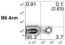
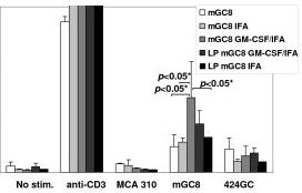
Tumor-specific IFN-γ release and cell-mediated cytotoxicity after vaccination with mGC8 cells and GM-CSF.

GM-CSF
  • Vaccination with mGC8 with or without LP, GM-CSF, and IFA.

Tumor cell vaccination (prophylactic/therapeutic), LRAST

  • To determine the immunogenicity of the tumor cells, 107 tumor cells were irradiated with 10,000 rad and subcutaneously injected into mice.
  • Two weeks later, the mice were challenged by subcutaneous injection of 3 × 10⁶ viable tumor cells into the opposite flank.
  • Experimental groups generally consisted of 5 mice.
  • Tumor development was followed by serial measurements of the tumor diameter and is depicted as tumor size (mm2) = d × D, where d and D were the shortest and the longest tumor diameter, respectively.
  • Animals were euthanized when D reached 10 mm.
  • Lymphopenia was induced by i.p.
  • injection of cyclophosphamide (200 mg/kg, , ).
  • This dose was chosen since earlier studies have shown an increased proliferation and long-term survival of antigen-specific T cells at this dose of cyclophosphamide, alone or in combination with fludarabine [7 naïve syngeneic splenocytes followed by s.c. vaccination with irradiated mGC8 cells (107, 10,000 rad) with or without a…
  • To determine the immunogenicity of the tumor cells, 107 tumor cells were irradiated with 10,000 rad and subcutaneously injected into mice.
  • Two weeks later, the mice were challenged by subcutaneous injection of 3 × 10⁶ viable tumor cells into the opposite flank.
  • Experimental groups generally consisted of 5 mice.
  • Tumor development was followed by serial measurements of the tumor diameter and is depicted as tumor size (mm2) = d × D, where d and D were the shortest and the longest tumor diameter, respectively.
  • Animals were euthanized when D reached 10 mm.
  • Lymphopenia was induced by i.p.
  • injection of cyclophosphamide (200 mg/kg, , ).
  • This dose was chosen since earlier studies have shown an increased proliferation and long-term survival of antigen-specific T cells at this dose of cyclophosphamide, alone or in combination with fludarabine [7 naïve syngeneic splenocytes followed by s.c. vaccination with irradiated mGC8 cells (107, 10,000 rad) with or without a s.c. injection of GM-CSF (1 μg, , ) diluted in HBSS and emulsified with an equal volume of incomplete Freund's adjuvant (IFA, Taufkirchen, ) as described elsewhere [6) were injected 4 days before vaccination and tumor vaccinations were repeated every two weeks for a total of 4 vaccinations.

Read more

BMDC culture

  • Bone marrow was extracted from the femur and tibia passed through a screen and washed in cold media.
  • 2×10⁶ bone marrow cells were resuspended in 10 ml of DC media (RPMI, 10% LPS free FCS, β-mercaptoethanol, L-glutamine, 40 ng/ml GM-CSF and cultured in 10 cm petri dishes at 37°C.
  • On day 3, 10 ml of DC media was added to the culture.
  • On day 6 and 8, 10 ml of culture was removed, resuspended in 10 ml of fresh DC media and added back to the culture.
  • On day 9, BMDCs were resuspended in DC media (ex GM-CSF) at 2×10⁶cell/ml, re-plated in 24 well plates and stimulated with LPS (100 ng/ml), Poly I∶C (100 µg/ml), and/or IFNα (1000 u/ml).
  • On day 10, BMDCs were pulsed for 2 hours with gp33 peptide alone or gp33, gp276 and gp61 peptides (10−6 M) respectively, and washed prior to use in in…
  • Bone marrow was extracted from the femur and tibia passed through a screen and washed in cold media.
  • 2×10⁶ bone marrow cells were resuspended in 10 ml of DC media (RPMI, 10% LPS free FCS, β-mercaptoethanol, L-glutamine, 40 ng/ml GM-CSF and cultured in 10 cm petri dishes at 37°C.
  • On day 3, 10 ml of DC media was added to the culture.
  • On day 6 and 8, 10 ml of culture was removed, resuspended in 10 ml of fresh DC media and added back to the culture.
  • On day 9, BMDCs were resuspended in DC media (ex GM-CSF) at 2×10⁶cell/ml, re-plated in 24 well plates and stimulated with LPS (100 ng/ml), Poly I∶C (100 µg/ml), and/or IFNα (1000 u/ml).
  • On day 10, BMDCs were pulsed for 2 hours with gp33 peptide alone or gp33, gp276 and gp61 peptides (10−6 M) respectively, and washed prior to use in in vitro proliferation assays or intravenous infusions into treated mice.
  • For intravenous infusions, 2×10⁶ BMDCs prepared in 200 µl of sterile HBSS were given to each treated mouse.
  • For in vivo treatment, mice were infused with 10000 u of IFNα.

Read more

Immunoprotection analysis of Com1- and HspB-pulsed BMDCs

  • Mouse bone marrow dendritic cells (BMDCs, CD11c+) were isolated from the bone marrow of BALB/c mice, according to the protocol described previously [2.
  • Approximately 1 × 10⁶ cells was added to each well of a six-well plate, and mouse GM-CSF (20 ng/ml) and IL-4 (10 ng/ml) was added to the culture medium every other day.
  • After 6 days of culture, BMDCs were stimulated with C.
  • burnetii I Ag (10 μg/ml), Com1 (10 μg/ml), HspB (10 μg/ml), E.
  • coli LPS (2 μg/ml), or 25 μl elution buffer (mock pulse) for 24 h at 37°C and 5% CO2.

Generation of BMDC and BMMC

  • Complete medium'>medium'>RPMI (cmedium'>medium'>RPMI), medium'>medium'>RPMI 1640 medium containing 10% fetal calf serum , was used as culture medium.
  • For BMDC induction, 5×10⁶ BM cells were cultured supplemented with 10 ng/ml recombinant murine GM-CSF for five days

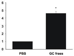
GC frass-induced PAR-2 mRNA and cytokine production from BMDC.

GM-CSF
  • Bone marrow was isolated from BALB/c mice and cultured in the presence of GM-CSF for 6 days.

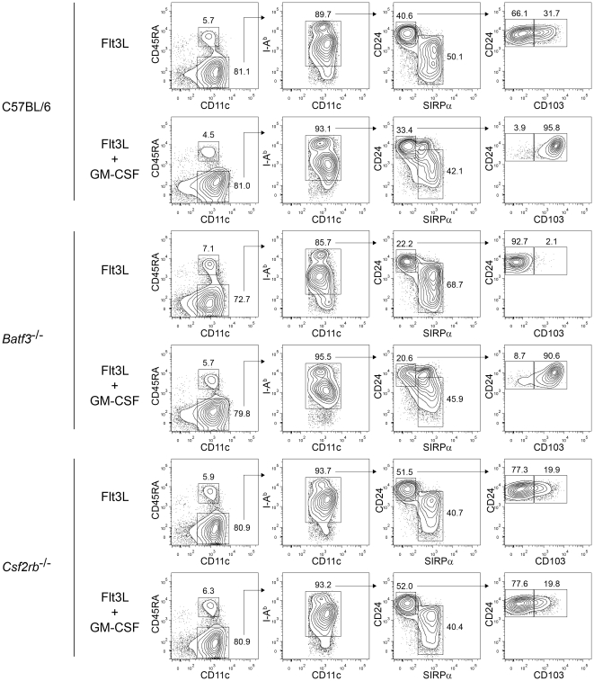
GM-CSF induces CD103 expression on Flt3L-derived CD8α-equivalent DCs.

murine GM-CSF
  • FACS analysis of developing DCs in bone marrow cells from C57BL/6, Batf3

Phagocytic cells assays

  • The monocyte/macrophage J774.3 cell line used in this study was kindly provided by Dr. María Inés Becker .
  • J774.3 cells were routinely grown in high-glucose medium'>DMEM medium (GIBCO), supplemented with 10% Fetal Bovine Serum and 1 mM HEPES (GIBCO) in T75 bottles.
  • Cells were incubated at 37°C and 5% CO2 until 95% of confluence.
  • Before infection assays, cells were treated with 0.1 mg/ml trypsine for 5 min, recovered in 50 ml polypropylene tubes, and centrifuged at 1,800× g for 5 min at room temperature.
  • After three washes with supplemented DMEM medium, cell number and viability was determined in a haemocytometer, using the trypan blue staining (1 mg/ml).
  • 5×10⁵ cells/ml were seeded in 24 well-plates and incubated overnight at 37°C and 5% CO2.
  • DCs were prepared from bone marrow precursors of C57BL/6 mice.
  • Cells were incubated in complete RPMI 1640 medium supplemented with 5% FCS…
  • The monocyte/macrophage J774.3 cell line used in this study was kindly provided by Dr. María Inés Becker .
  • J774.3 cells were routinely grown in high-glucose medium'>DMEM medium (GIBCO), supplemented with 10% Fetal Bovine Serum and 1 mM HEPES (GIBCO) in T75 bottles.
  • Cells were incubated at 37°C and 5% CO2 until 95% of confluence.
  • Before infection assays, cells were treated with 0.1 mg/ml trypsine for 5 min, recovered in 50 ml polypropylene tubes, and centrifuged at 1,800× g for 5 min at room temperature.
  • After three washes with supplemented DMEM medium, cell number and viability was determined in a haemocytometer, using the trypan blue staining (1 mg/ml).
  • 5×10⁵ cells/ml were seeded in 24 well-plates and incubated overnight at 37°C and 5% CO2.
  • DCs were prepared from bone marrow precursors of C57BL/6 mice.
  • Cells were incubated in complete RPMI 1640 medium supplemented with 5% FCS , 2 mM glutamine, 1 mM non-essential amino acids, 1 mM pyruvate, 1 mM HEPES, and 10 ng/ml of recombinant murine GM-CSF .
  • All cell culture media were acquired from GIBCO .
  • Culture media was replaced every 2 days.
  • After 6 days, the phenotype of DCs was analyzed by flow cytometry for the expression of the surface markers CD11c, CD86 and CD40, which revealed over 70% CD11c+ with an immature phenotype.
  • Before the infection assays, DCs were washed three times with PBS and then culture media was replaced with complete medium'>RPMI medium without antibiotics.
  • DCs and macrophages were infected with S. Enteritidis PT1 at a multiplicity of infection (MOI) equal to 25.
  • The MOI was confirmed by plating serial dilutions of bacterial cultures on LB agar.
  • After 1 h of incubation at 37°C and 5% CO2, the supernatant of infected cells was recovered and stored for genomic DNA preparation.
  • Cells were washed two times with PBS and 1 ml of appropriate medium supplemented with gentamicin 50 mg/ml was added to cell cultures to kill the remaining extracellular bacteria.
  • After 2, 18 and 24 h of infection, cells were removed from the wells and centrifuged at 783× g for 5 min.
  • No significant changes on cell viability were observed after infection with S. Enteritidis at the time points used on the experiments (data not shown).
  • To recover intracellular bacteria, phagocytic cells were treated with 1 ml of lysis solution (19% ethanol, 0.1% SDS, 1% saturated basic phenol) for 30 min on ice.
  • After the incubation period, the cell lysate was centrifuged at 7,043× g for 5 min, and genomic DNA was extracted following the methodology mentioned above.
  • In order to quantify the amount of intracellular bacteria at different time points, either 100,000 DCs or 10,000 J774.3 cells were treated with 1 ml of PBS-0.1% triton X-100 for 15 min at room temperature.
  • 100 µl of cell lysates were plated on LB agar and the plates were incubated at 37°C for 18 h. In parallel, an equal amount of bacteria used to infect cells was incubated in 1 ml of cell medium.
  • The incubation was performed for 2 h and after that time bacteria were recovered by centrifugation at 7.043× g for 5 min, and genomic DNA was prepared as described above.

Read more

Generation of bone marrow-derived DCs

  • Mouse BMDCs were generated from bone marrow progenitor cells, according to the modified protocol of[