Human SDF-1 alpha Recombinant
Categories: Recombinant Human CytokinesCXC chemokines$70.00 – $3,500.00
Description
Accession
P48061
Source
Optimized DNA sequence encoding Human Stromal Cell-Derived Factor-1 alpha mature chain was expressed in Escherichia Coli.
Molecular weight
Native human SDF-1 alpha, generated by the proteolytic removal of the signal peptide and propeptide,the molecule has a calculated molecular mass of approximately8 kDa. Recombinant SDF-1 alpha is a monomer protein consisting of69 amino acid residue subunits, migrates as an approximately8 kDa protein under non-reducing and reducing conditions in SDS-PAGE.
Purity
>97%, as determined by SDS-PAGE and HPLC
Biological Activity
Determined by its ability to chemoattract human U937 expressing CXCR4 Determined by calcium flux with human U937 cells.
Protein Sequence
MNAKVVVVLV LVLTALCLSD GKPVSLSYRC PCRFFESHVA RANVKHLKIL NTPNCALQIV ARLKNNNRQV CIDPKLKWIQ EYLEKALNK R FKM
Endotoxin
Endotoxin content was assayed using a LAL gel clot method. Endotoxin level was found to be less than 0.1 ng/µg(1EU/µg).
Presentation
Recombinant SDF-1 alpha was lyophilized from a 0.2μm filtered PBS, pH.4.
Reconstitution
A quick spin of the vial followed by reconstitution in distilled water to a concentration not less than 0.1 mg/mL. This solution can then be diluted into other buffers.
Storage
The lyophilized protein is stable for at least years from date of receipt at -20° C. Upon reconstitution, this cytokine can be stored in working aliquots at2° -8° C for one month, or at -20° C for six months, with a carrier protein without detectable loss of activity. Avoid repeated freeze/thaw cycles.
Usage
This cytokine product is for research purposes only.It may not be used for therapeutics or diagnostic purposes.
Interactor
P15172
Biological Process
Molecular function
Molecular function
Methods
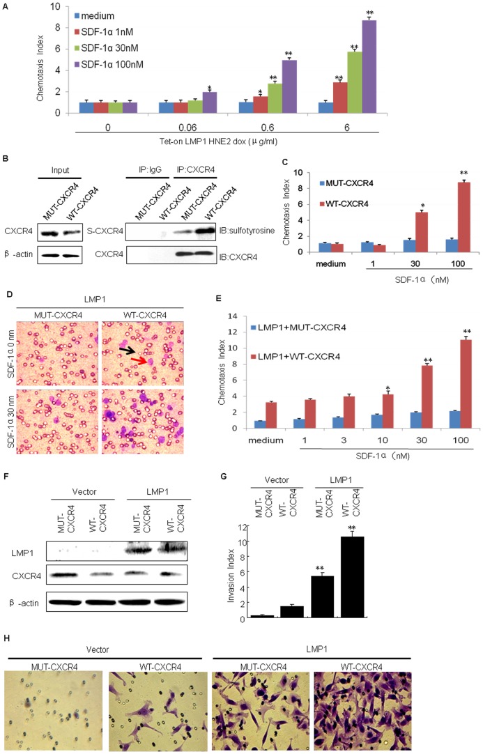
LMP1 up-regulates cellular chemotactic activity and invasiveness through the tyrosine sulfation of CXCR4.
- The migration of Tet-on-LMP1 HNE2 cells in response to SDF-1α was measured using chemotaxis chambers.
Materials
- Mouse anti-His monoclonal antibody was bought from .
- Complete EDTA-free protease inhibitor cocktail tablets were bought from.
- The commercial recombinant human chemokines CCL11, CCL24 and CXCL12 were bought from .
Chemotaxis assay
- A migration assay was performed in 48- or 96-well Corning Costar transwell chambers with porous polycarbonate membranes with a pore size of 5 um .
- hMSC at passage 4 were resuspended at 1 × 10⁶/mL in the migration medium supplemented with 1% BSA and insulin transferrin selenium (ITS) and seeded in the upper chamber.
- The following human recombinant proteins were used as chemoattractants in the lower compartment: hepatocyte growth factor (HGF), platelet-derived growth factor-AB (PDGF-AB), epidermal growth factor (EGF), VEGF-121, basic fibroblast growth factor (bFGF), insulin-like growth factor (IGF-1), macrophage inflammatory protein-3β (MIP-3β), macrophage inflammatory protein-1α (MIP-1α), B cell attracting chemokine-1 (BCA-1), regulation upon activation normal T cell express sequence (RANTES), growth regulated proteinα (GROα), fractalkine, SDF-1α, IL-1β, IL-6, IL-8, TNF-α .
- The concentration of each cytokine was optimized to ensure maximal cell migration through the porous membrane.
- The concentrations were: 40 ng/mL HGF, 10 ng/mL PDGF-AB, 10 ng/mL EGF, 10 ng/mL VEGF-121,…
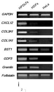
Results of RT-PCR and qRT-PCR.
- RT-PCR of expression of CXCL12 and connective tissue specific markers.
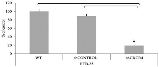
The confirmation of CXCR4 gene downregulation.
- The expression of phospho-AKT and phospho-MAPK after human SDF-1β (100 ng/ml) stimulation of control cells (HTB-35 WT and HTB-35 shCONTROL) and examined the cells (HTB-35 shCXCR4) at 2, 5, 10 and 30 min.
Cell Culture and Treatments
- In experiments investigating CXC-chemokine signaling, cells were incubated in serum-free, phenol-red free RMPI 1640 or medium'>DMEM medium for 16h prior to exposure to 3nM recombinant human (rh)-CXCL8, 100ng/ml rh-CXCL12 or 100ng/ml rh-CCL2 .
- For inhibition of chemokine signaling, cells were treated with RS102895 (CCR2 antagonist, 10nM, , ), AMD3100 (CXCR4 antagonist, 25μg/ml) or a CXCR1/2-derived pepducin (x1/2pal-i3, 300nM, , ).
- Control cells were treated with the appropriate vehicle or NT-pepducin (x1/2pal-con) sequence as required.
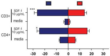
Migration of human T lymphocytes in response to SDF-1 A).
- Percentage of human T lymphocytes migrating towards or away from SDF-1 (10 μg mL−1) and media (control) Significantly greater numbers of T cells (both pooled CD3+ and CD4+ cells) migrate away from SDF-1 than towards (p<0.05, Student's t test).
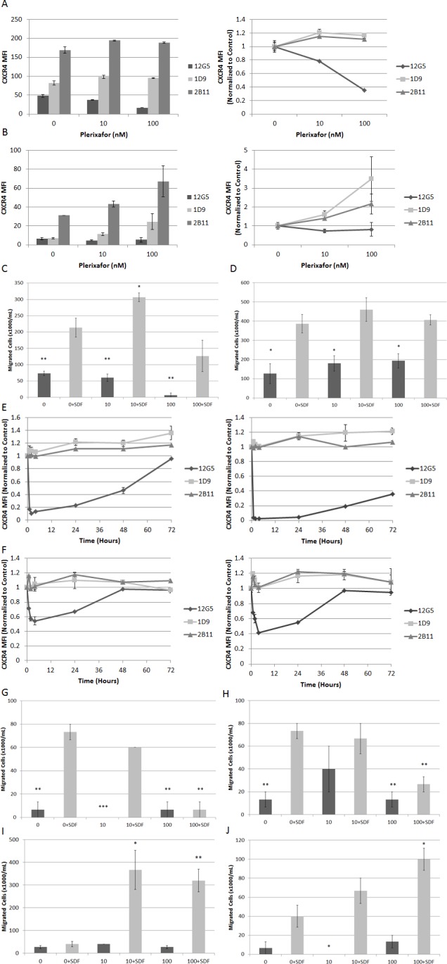
Extended treatment with plerixafor modulates surface expression of surface CXCR4 and affects SDF-1α-induced chemotaxis Cell lines were treated with plerixafor (10 nM, 100 nM) or vehicle control (0 nM) for 72 hours and analyzed by flow cytometry and chemotaxis assays.
- Chemotaxis toward medium with or without recombinant SDF-1α (150 ng/mL) after 24 hours in Nalm-6 and HB-1119.
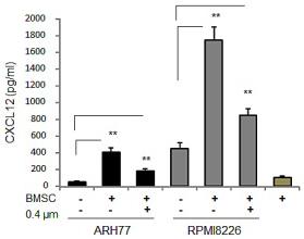
Both MM and BMSC cells express CXCL12.
- Interaction with BMSCs elevates CXCL12 expression in MM cell lines Secretion of CXCL12 by MM cell lines RPMI8226 and ARH77, incubated in the absence or presence of BMSCs, in direct contact or separated by 0.4 μm-transwells for 48 hours, evaluated by ELISA.