Human Interleukin-1 alpha Recombinant
Categories: Interleukin-1 familyRecombinant Human Cytokines$70.00 – $4,700.00
Description
Accession
P01583
Source
Optimized DNA sequence encoding Human Interleukin-1 alpha mature chain was expressed in Escherichia Coli.
Molecular weight
Mature human IL-1 alpha, is generated by the proteolytic removal of the signal peptideand propeptide. The molecule has a calculated molecular mass of approximately kDa. Recombinant Interleukin-1a is a monomer protein consisting of160 amino acid residue subunits and migrates as an approximately18 kDa protein under non-reducing conditions and reducing conditions in SDS-PAGE.
Purity
>97%, as determined by SDS-PAGE and HPLC
Biological Activity
The ED(50) was determined by thedose-dependent stimulation of murine D10S cells is ≤.002 ng/ml, corresponding to a specific activity of ≥2 x units/mg.
Protein Sequence
MAKVPDMFED LKNCYSENEE DSSSIDHLSL NQKSFYHVSY GPLHEGCMDQ SVSLSISETS KTSKLTFKES MVVVATNGKV LKKRRLSLSQ SITDDDLEAI ANDSEEEIIK PRSAPFSFLS NVKYNFMRII KYEFILNDAL NQSIIRANDQ YLTAAALHNL DEAVKFDMGA YKSSKDDAKI TVILRISKTQ LYVTAQDEDQ PVLLKEMPEI PKTITGSETN LLFFWETHGT KNYFTSVAHP NLFIATKQDY WVCLAGGPPS ITDFQILENQ A
Endotoxin
Endotoxin content was assayed using a LAL gel clot method. Endotoxin level was found to be less than 0.1 ng/µg(1EU/µg).
Presentation
Interleukin-1 alphawas lyophilized from a 0.2 μm filtered PBS solution pH7.0.
Reconstitution
A quick spin of the vial followed by reconstitution in distilled water to a concentration not less than 0.1 mg/mL. This solution can then be diluted into other buffers.
Storage
The lyophilized protein is stable for at least years from date of receipt at -20° C. Upon reconstitution, this cytokine can be stored in working aliquots at2° -8° C for one month, or at -20° C for six months, with a carrier protein without detectable loss of activity. Avoid repeated freeze/thaw cycles.
Usage
This cytokine product is for research purposes only.It may not be used for therapeutics or diagnostic purposes.
Biological Process
Molecular function
Molecular function
Molecular function
Methods
Size separation of proteins in CM of PDT-treated T-Ep.
- Detection of the FSA activity by chromatographed COS-1 cell-derived IL-1α is included in (B and C).
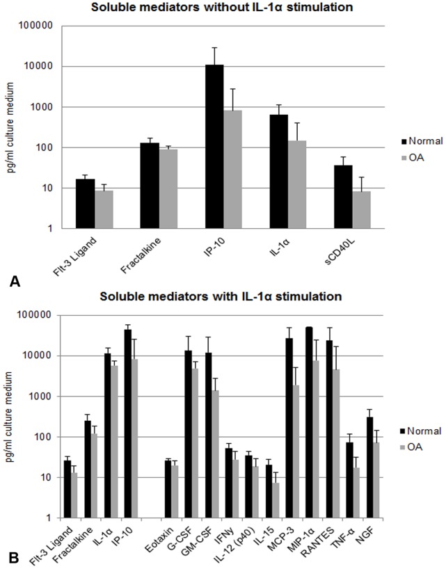
Soluble mediator secretion by normal and OA synovial tissue explants (STEs) with or without IL-1α.
- Graphs demonstrate all soluble mediators which were significantly (p<0.01) different between A. normal (black bars) and OA (grey bars) STEs and B. normal and OA STEs under pro-inflammatory (IL-1α) conditions.
Cell Culture
- VSMCs, EL4, HEK, and HeLa cells were cultured in DMEM and Jurkat and THP-1 cells in RPMI 1640, all supplemented with penicillin, streptomycin, L-glutamine, and 10% FCS.
- Human monocyte-derived macrophages were differentiated as described previously (2, clarified, and stored at −80°C.
- Cells were also made necrotic by incubation with 7-BIO (25 μM) or digitonin (0.1%) (data not shown) or by overnight hypoxic exposure.
- To activate inflammasomes, cells were treated with LPS (1 μg/ml; 4 hr), followed by ATP (5 mM) or Nigericin (20 μM) for 30 min.
- Calpain activity was determined with Calpain-Glo .
- VSMCs, HEK, and THP-1 cells were transfected with pcDNA3 with nucleofection or FugeneHD .
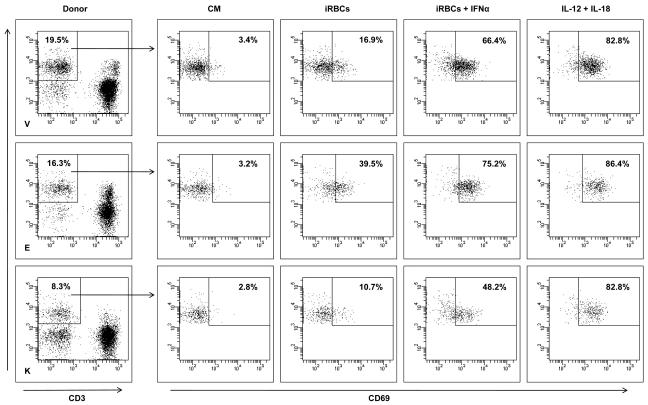
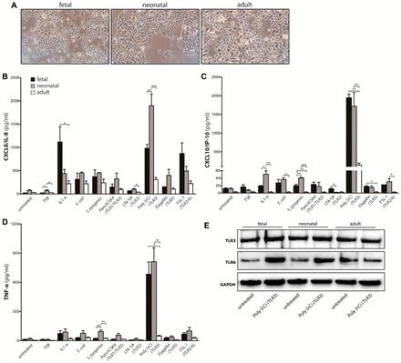
Bacterial stimuli and TLR ligands induce distinct immune responses in fetal, neonatal and adult keratinocytes.
- IL1α was used as positive control, cell culture medium (untreated) and TSB as negative controls.
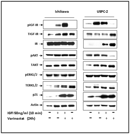
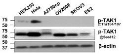
TAK1 exerts its functional effects through activation of NF -κB pathways by phosphorylation at Ser412 (A) Western blot analysis demonstrated that p-TAK1 at Thr184/187 was just found in HEK293 and Hela cells only, while p-TAK1 at Ser412 was generally found in all cell types including ovarian cancer cell lines (n=4).
- After 48 hours, Human IL-1α (10ng/ml) was used to treat the transfected cells with various time points.