Human Interferon-alpha 2a Recombinant
Categories: Interferon-IL10 familyRecombinant Human Cytokines$70.00 – $880.00
Description
Accession
P01563
Source
Optimized DNA sequence encoding Human Interferon-alpha mature chain was expressed in Escherichia Coli.
Molecular weight
Recombinant human IFN-alphaa, generated by the proteolytic removal of the signal peptide and propeptide, and has a calculated molecular mass of approximately 19 kDa. Recombinant Interferon alphaais a monomeric protein consisting of 165 amino acid residue subunits, and migrates as an approximately 19 kDa protein under non-reducing conditions and reducing conditions in SDS-PAGE.
Purity
>98%, as determined by SDS-PAGE and HPLC
Biological Activity
Theactivity was determined bya viral resistance assay of Human WISH cells, andwas found to be in the range ofx108 IU/mg.
Protein Sequence
MALTFALLVA LLVLSCKSSC SVGCDLPQTH SLGSRRTLML LAQMRKISLF SCLKDRHDFG FPQEEFGNQF QKAETIPVLH EMIQQIFNLF STKDSSAAWD ETLLDKFYTE LYQQLNDLEA CVIQGVGVTE TPLMKEDSIL AVRKYFQRIT LYLKEKKYSP CAWEVVRAEI MRSFSLSTNL QESLRSKE
Endotoxin
Endotoxin content was assayed using a LAL gel clot method. Endotoxin level was found to be less than 0.1 ng/µg(1EU/µg).
Presentation
Recombinant Interferon alphaawas lyophilized from a 0.2 μm filtered PBS solution pH7.0.
Reconstitution
A quick spin of the vial followed by reconstitution in distilled water to a concentration not less than 0.1 mg/mL. This solution can then be diluted into other buffers.
Storage
The lyophilized protein is stable for at least years from date of receipt at -20° C. Upon reconstitution, this cytokine can be stored in working aliquots at2° -8° C for one month, or at -20° C for six months, with a carrier protein without detectable loss of activity. Avoid repeated freeze/thaw cycles.
Usage
This cytokine product is for research purposes only.It may not be used for therapeutics or diagnostic purposes.
Interactor
P17181
Interactor
Biological Process
Molecular function
Methods
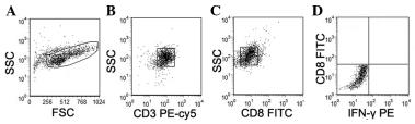
IFN-γ Stimulation Assay
- Human IFN-γ at 10 ng ml−1 was added to day 30 thymic differentiation cultures in BEL medium.
- After 72 hr of IFN-γ stimulation, differentiated cells were harvested and analyzed for HLA-DR expression by flow cytometry.
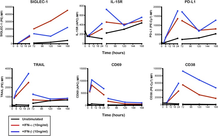
Time-course of the expression of six IFN-inducible proteins on the surface of CD14+ monocytes.
- PBMCs isolated from a healthy donor were cultured with IFN-α (10 ng/mL; red line), IFN-β (10 ng/mL; blue line), or culture medium (unstimulated; black line).
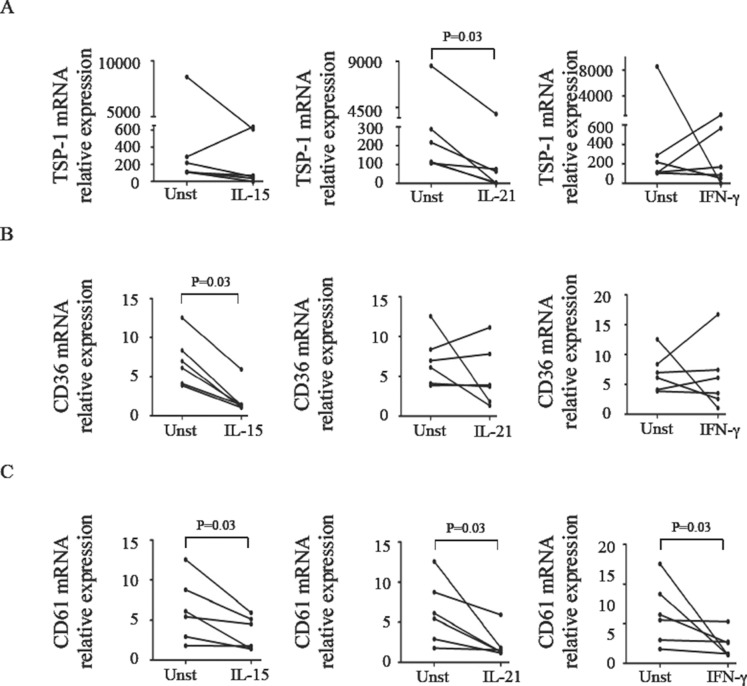
IL-15, IL-21 and IFN-γ modulate expression of TSP-1, CD36, and CD61 in normal LPMC.
- LPMC isolated from the jejunal mucosa of 6 normal controls were cultured with or without (Unst = unstimulated) IL-15 (50 ng/ml), IL-21 (50 ng/ml) and IFN-γ (100 ng/ml) and then analyzed for TSP-1 , CD36 , and CD61 RNA expression by Real-Time PCR and levels were normalized to β-actin.
GTS-21 reduces the percentages of IFN-γ+ T cells in RA CD4+ T cells during Th1 differentiation.
- Percentages of IFN-γ+ cells among CD3+CD8− T cells stimulated by anti-CD3/-CD28 for 72 h. Percentages of IFN-γ+ cells among CD3+CD8− T cells under Th1-differentiation conditions for 72 h. (G and H) Effect of GTS-21 alone or with αBgt on the expression of IFN-γ by CD3+CD8− T cells from patients with RA.
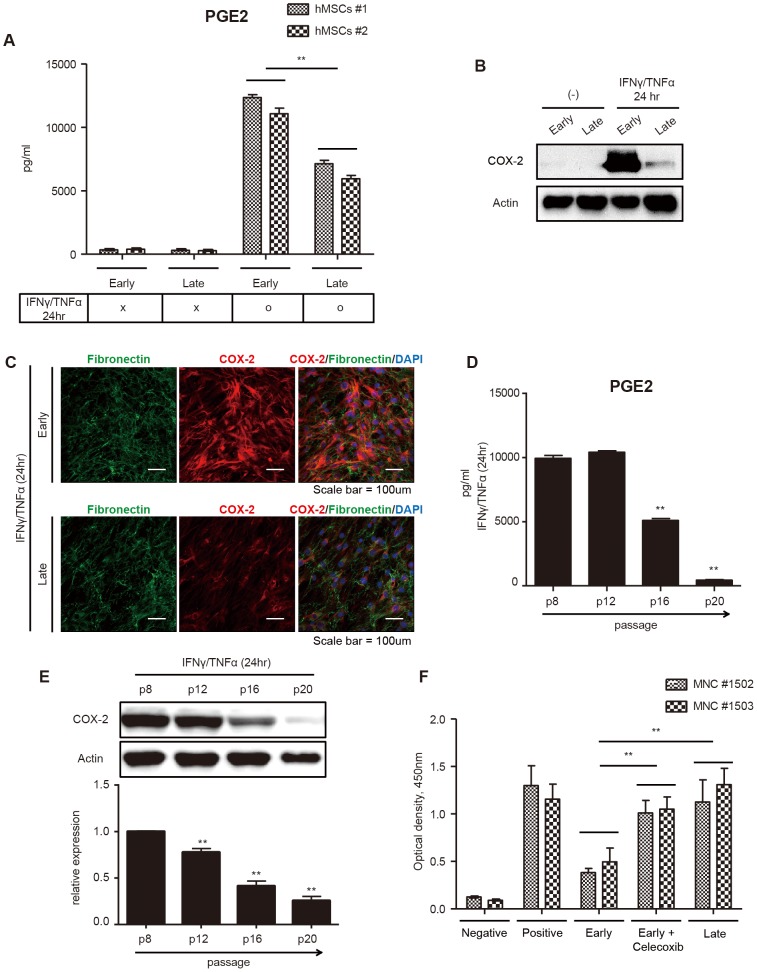
Declined immune-inhibitory effect of late-passage hMSCs is regulated by PGE2 and COX-2.
- Early- and late-passage hMSCs were treated with or without IFN-γ and TNF-α for 24 hours.
Declined immune-inhibitory effect of late-passage hMSCs is regulated by PGE2 and COX-2.
- Early- and late-passage hMSCs were treated with or without IFN-γ and TNF-α for 24 hours.
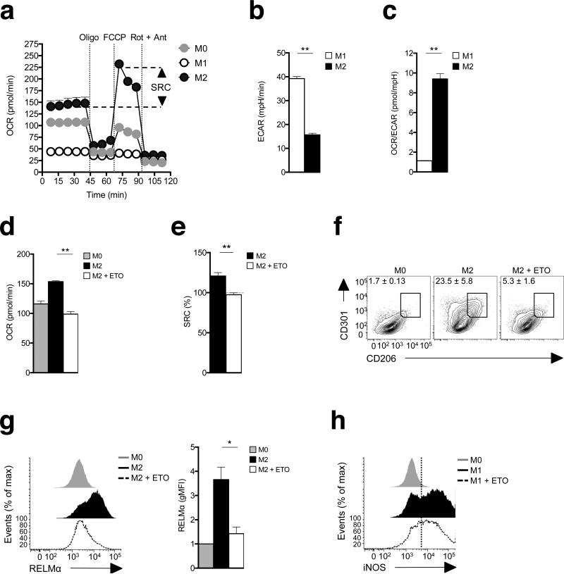
M2 activation is marked by increased spare respiratory capacity and is dependent on fatty acid oxidation.
- (a) Bone marrow-derived macrophages were cultured in medium without (M0) or with IFN-γ + LPS (M1), or with IL-4 (M2) for 24 h and then oxygen consumption rates was determined using an XF-96 Extracellular Flux Analyzer during sequential treatments with oligomycin, FCCP, and rotenone/antimycin.
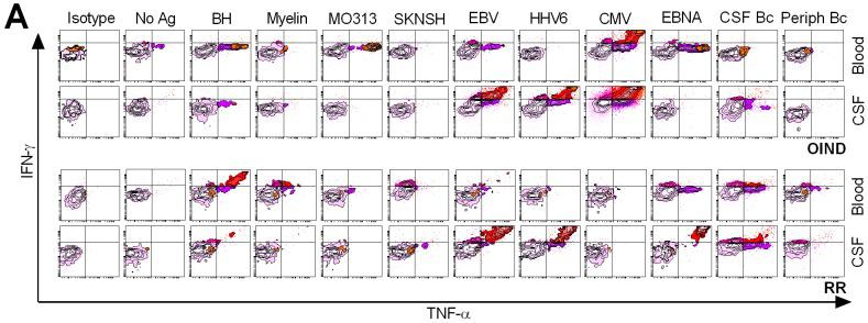
Differential phenotypes of peripheral and intrathecal CD4+ T cells in response to foreign Ag's.
- FACS plots illustrate intracellular TNF-α and IFN-γ secretion by peripheral (Blood) and intrathecal CD4+ T cells in response to all candidate Ag's.
Monocytes stimulated in vitro with B. malayi Mf lysate develop a specific activation phenotype.
- Monocytes were left unstimulated or stimulated for 24 h with 100 ng/ml LPS+20 ng/ml IFN-γ, 20 ng/ml IL-4 or 20 µg/ml B. malayi Mf lysate.
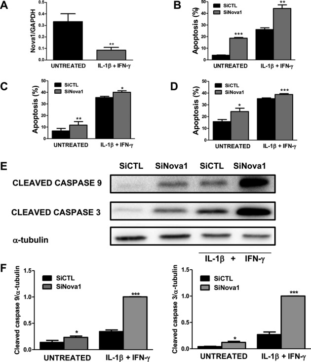
Nova1 KD increases apoptosis under basal condition and following cytokine treatment.
- Primary rat beta cells were exposed to the pro-inflammatory cytokines IL-1β + IFN-γ for 48 h and then collected for mRNA expression analyses.

