Human IL-6 Receptor Recombinant (IL-6R)
Category: Surface & Soluble Receptors$160.00 – $2,200.00
Description
Accession
P08887
Source
Optimized DNA sequence encoding extracellular domain of human IL-6 receptor (CD126) including a C-terminal PolyHis tag was expressed in HEK293 cells.
Molecular weight
Recombinant IL-6 receptor (CD126) is a monomer protein consisting of357 amino acid residue subunits,due to glycosylation migrates as an approximately60-65 kDa protein on SDS-PAGE.
Purity
>97%, as determined by SDS-PAGE and HPLC
Biological Activity
Recombinant IL-6R (CD126) activity was determined by its ability to enhance the IL6 activity on M1 mouse myeloid leukemia cells. The ED50 for this effect is typically 20-80 ng/ml.
Endotoxin
Endotoxin content was assayed using a LAL gel clot method. Endotoxin level was found to be less than 0.1 ng/µg(1EU/µg).
Presentation
Recombinant Human Interleukin-6 receptor (CD126) is supplied as a 0.2 μm filtered PBS solution,pH7.2.
Storage
Recombinant human IL-6R, as supplied, can be stored in working aliquots at 2° - 8° C for one month, or at -20°C to-70°Cfor twelve months. Avoid repeated freeze/thaw cycles.
Usage
This product is for research purposes only.It may not be used for therapeutics or diagnostic purposes.
Interactor
Interactor
Molecular function
Methods
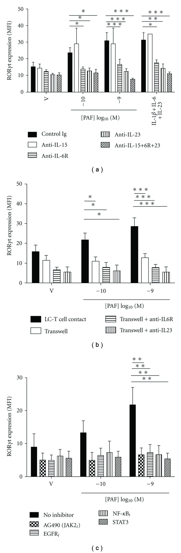
PAF-stimulated Th17 development is dependent on cytokines, LC-T cell contact and selected signaling pathways.
- (a) Monocyte-derived LC were stimulated with either vehicle or PAF in the presence of neutralizing Ab to IL-15, IL-6R, and/or IL-23, or control Ig, and cocultured with antiCD3/CD28-activated CD4+ T cells for 5 days.
Cell Culture
- Frozen female CD34+ CBCs were supplied by Bio-Resource Center .
- C34+ CBCs were cultured in hematopoietic culture medium [serum-free X-Vivo10 containing 50 ng/mL IL-6 , 50 ng/mL sIL-6 , 50 ng/mL SCF , ten ng/mL TPO , and 20 ng/mL Flt3/4 ligand ].
- Reprogrammed cells were cultured in feeder-less primate ES cell medium Repro FF (, .
- No. RCHEMD004), ReproFF2 (ReproCELL, cat No. RCHEMD006), mTeSR1 ( catalog number 05850) or E8 (16) supplemented with five ng/mL bFGF (total bFGF ten ng/mL) on Pronectin F-coated dishes.
- Passage of human iPSCs was previously described
Cell proliferation assay
- HEK293T cells were transfected with mc_mock, mc_sTNFR-Fc, or mc_anti-IL-6R and the conditioned media were collected 24 h post-transfection.
- A-FLS were incubated with or without human IL-6 (100 ng/mL& , , ), sIL-6 (100 ng/mLocky , ), and TNFα (20 ng/mL& ) for 72 h. uring the incubationA-FLS were treated with the conditioned media of HEK293T cells transfected with mc_mock, mc_sTNF-Fc, mc_anti-IL-6 or PBS (as a negative control).
- Cell proliferation was assessed using the cell counting kit-8 (CCK-8 , , ), according to the manufacturer's instructions.
Enzyme-linked immunosorbent assay (ELISA)
- To analyze the amounts of anti-IL-6R antibody and sTNFR2-Fc protein expressed by the transfected cells, the culture media of HEK293T cells transfected with the indicated minicircle vectors were analyzed by ELISA at 24 h post-transfection.
- The levels of sTNFR2-Fc were quantified using human sTNF-R (80 kDa) Platinum ELISA (eBioscience, San Diego, CA), according to the manufacturer's instructions.
- ELISA was performed to determine the levels of anti-IL-6R antibodies.
- Briefly, a 96-well microtiter plate was coated with commercial anti-human IL-6R antibodies (B-R6; eBioscience) at a concentration of 0.5 μg/mL in coating buffer, and incubated overnight at 4°C.
- The plate was washed five times with washing buffer, and incubated with 1 μg/mL of human sIL-6R at room temperature (RT) for 1 h. After washing, the plate was incubated with blocking solution for 1 h at RT, followed by incubation with 100 μL of serially diluted tocilizumab (used as a standard), and the conditioned media from HEK293T cells were transfected…

