Human Glial-Derived Neurotrophic Factor Recombinant
Categories: Neurotrophic factorsRecombinant Human Cytokines$70.00 – $4,700.00
Description
Accession
P39905
Source
Optimized DNA sequence encoding Human GDNF mature chain was expressed in Escherichia Coli.
Molecular weight
Native Human Glial-Derived Neurotrophic Factor is generated by the proteolytic removal of the signal peptide and propeptide, the molecule has a calculated molecular mass of approximately 15 kDa. Recombinant GDNF is a disulfide-linked homodimeric protein consisting of two 125 amino acid residue subunits,and migrates as an approximately 30 kDa protein under non-reducing conditions and as 15 kDa under reducing conditions in SDS-PAGE.
Purity
>95%, as determined by SDS-PAGE and HPLC
Biological Activity
The ED(50) was determined by theproliferation of rat C6 cells is <.1 ng/ml, corresponding to a specific activity of > x units/mg.
Protein Sequence
MKLWDVVAVC LVLLHTASAF PLPAGKRPPE APAEDRSLGR RRAPFALSSD SNMPEDYPDQ FDDVMDFIQA TIKRLKRSPD KQMAVLPRRE RNRQAAAANP ENSRGKGRRG QRGKNRGCVL TAIHLNVTDL GLGYETKEEL IFRYCSGSCD AAETTYDKIL KNLSRNRRLV SDKVGQACCR PIAFDDDLSF LDDNLVYHIL RKHSAKRCGC I
Endotoxin
Endotoxin content was assayed using a LAL gel clot method. Endotoxin level was found to be less than 0.1 ng/µg(1EU/µg).
Presentation
RecombinantGlial-Derived Neurotrophic Factor was lyophilized from a 0.2 μm filtered solution in.5% glycine,.5% sucrose,.01% Tween80, mM Glutamic acid, pH.5.
Reconstitution
A quick spin of the vial followed by reconstitution in distilled water to a concentration not less than 0.1 mg/mL. This solution can then be diluted into other buffers.
Storage
The lyophilized protein is stable for at least years from date of receipt at -20° C. Upon reconstitution, this cytokine can be stored in working aliquots at2° -8° C for one month, or at -20° C for six months, with a carrier protein without detectable loss of activity. Avoid repeated freeze/thaw cycles.
Usage
This cytokine product is for research purposes only.It may not be used for therapeutics or diagnostic purposes.
Interactor
Q8N5Y2
Interactor
Molecular function
Methods
Overexpression of EpICD in ROSA GS cells.
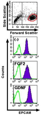
- Upregulation of EPCAM by GDNF stimulation.
Surface Plasmon resonance (Biacore) measurements
- MCer1 was immobilized on a CM5 chip in BIAcore 2000 with an amine group.
- Binding of Cer1 to hBMP2 , hBMP4, or hGNF (1 µg/ml& ) was analysed in triplicate at 25°C in HBS-P buffer supplemented with 10 mM HEPES, 150 mM NaCl and 0.005% Tween 20 at pH 7.4, with a flow rate of 20 µg/ml min.
- The kinetics and the dissociation constant (KD) were calculated with BIAevaluation software ver.
- 4.1 .
Preparation of Primary Hippocampal Cultures
- Primary hippocampal cultures were prepared from wild-type neonatal (E19) rat embryos (timed pregnant Sprague Dawley rats were obtained from Charles River Laboratories, Wilmington, MA) as described previously
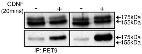
RET51 internalizes more efficiently than RET9.
- Retinoic acid–treated SH-SY5Y cells were serum starved, incubated with or without 100 ng/ml GDNF for 20 min, and lysed.
RET51 internalizes more efficiently than RET9.
- Retinoic acid–treated SH-SY5Y cells were serum starved, incubated with or without 100 ng/ml GDNF for 20 min, and lysed.
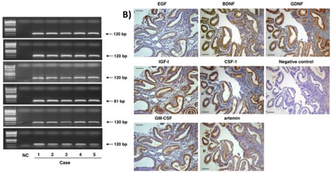
Expression of key growth factors in human endometrium.
- Brown signals for growth factors (EGF, IGF-1, GM-CSF, BDNF, CSF-1, artemin, and GDNF) were found following staining with specific antibodies.
Cell Culture and Biochemical Assays
- For DmRet phosphorylation assays, cells stably expressing DmRet-3xFLAG or DmRet-3xFLAG and V5-DmGfrlA were plated in 6-well plates at ∼12 M cells/well, in serum-free M3-BPYE medium.
- Expression was induced with 600 µM CuSO4 for 2 to 4 hours.
- Recombinant human GDNF was added at 50 ng/ml for 1 hour after which the cells were triturated, washed in PBS, and lysed in membrane lysis buffer supplemented with 1 mM Na2VO4.
- Insoluble material was sedimented by centrifugation and to the supernatants 0.5 µg of affinity-purified DmRet antibody was added.
- After 15-minute incubation on ice, protein A-sepharose (GE Healthcare) was added and the samples were rotated in cold for 1–2 hours.
- The precipitates were washed with the lysis buffer three times, eluted with Laemmli sample buffer, and run on 8% SDS-PAGE.
- The immunoblots were probed with anti-phosphotyrosine (4G10) and anti-FLAG (M2, Sigma) antibodies.
- For mammalian RET phosphorylation assay MG87RET cells that…
FGSC Culture and Growth Assay
- To evaluate the growth-promoting activity of each candidate feeder line on the FGSC cultures, 2×10⁵ cells of each feeder line were seeded into individual well of a 12-well plate in triplicate.
- The candidate feeder cells were irradiated for 8 Krad to generate growth-arrested feeder cells.
- The FGSCs isolated from ovaries of 18
Tg(ziwi:neo);Tg(ziwi:) transgenic fish (10–12 weeks old) using a published
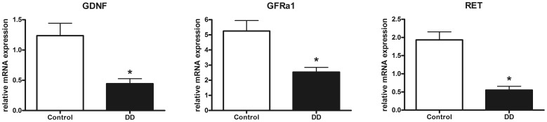
mRNA expression of the GDNF system in the muscularis propria of the human colon.
- mRNA expression of GDNF and its receptors GFRα1 and RET is significantly down-regulated in patients with DD compared to controls.