Human Bone Morphogenetic protein-4 Recombinant
Categories: Recombinant Human CytokinesTGF-beta familyTGF-beta family$70.00 – $3,550.00
Description
Accession
P12644
Source
Optimized DNA sequence encoding Human Bone Morphogenetic protein-4 mature chain was expressed in Escherichia Coli.
Molecular weight
Recombinant BMP-4 is a homodimeric protein consisting of 2x10⁶ amino acid residue subunits, and migrates as an approximately as a 12 kDa protein under reducing conditions in SDS-PAGE.
Purity
>95%, as determined by SDS-PAGE and HPLC
Biological Activity
The ED(50) was determined by its ability to induce alkaline phosphatase productionin mouse ATDC5 chondrogenic and was found to be in the range of 0.1-0.2 ug/ml.
Endotoxin
Endotoxin content was assayed using a LAL gel clot method. Endotoxin level was found to be less than 0.1 ng/µg(1EU/µg).
Presentation
Recombinant BMP-4 was lyophilized from a 0.2 μm filtered PBS, pH 7.2
Reconstitution
A quick spin of the vial followed by reconstitution in distilled water to a concentration not less than 0.1 mg/mL. This solution can then be diluted into other buffers.
Storage
The lyophilized protein is stable for at least years from date of receipt at -20° C. Upon reconstitution, this cytokine can be stored in working aliquots at 2° -8° C for one month, or at -20° C for six months, with a carrier protein without detectable loss of activity. Avoid repeated freeze/thaw cycles.
Usage
This cytokine product is for research purposes only.It may not be used for therapeutics or diagnostic purposes.
Interactor
O88273
Interactor
Interactor
Interactor
Interactor
Q05438
Interactor
Biological Process
Biological Process
Biological Process
Molecular function
Molecular function
Molecular function
Methods
Isolation, culture and 48 h induction of neural stem cells
- For 48 h induction using serum-free conditions with growth factors, dissociated neurospheres were cultured with recombinant human (rh) BMP4 250 ng/mL , rh Activin A 100 ng/mL and rh bFGF 100 ng/mL individually as well as in combinations as indicated in Supporting Information Fig S1 and
Growth Factors
- ecombinant human Noggin/Fc chimera and neutralizing anti-human /4 antibody from& , and from.
Growth Factors
- ecombinant human Noggin/Fc chimera and neutralizing anti-human /4 antibody from& , and from.
Growth Factors
- ecombinant human Noggin/Fc chimera and neutralizing anti-human /4 antibody from& , and from.
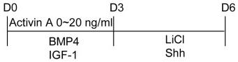
Effects of Activin A on paraxial mesodermal differentiation of mouse iPS cells.
- The cultures also contained BMP4 (10 ng/ml), IGF-1 (10 ng/ml), LiCl (5 mM), and Shh (10 ng/ml).
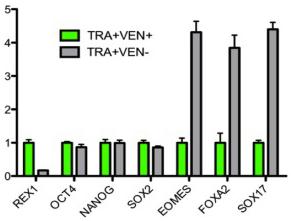
Loss of REX1 within the pluripotent population primes cells for differentiation.
- n = 3 E) Fold enrichment of the percentage of GATA4 positive endoderm cells generated from TRA+VEN− cells relative to those from TRA+VEN+ population after 3 days of treatment with Activin A and BMP4 in low serum media, as observed by immunocytochemistry.
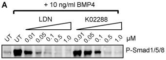
K02288 selectively inhibits BMP signaling.
- K02288 and LDN-193189 inhibited BMP4 induced Smad1/5/8 phosphorylation in C2C12 cells.
K02288 selectively inhibits BMP signaling.
- K02288 and LDN-193189 inhibited BMP4 induced Smad1/5/8 phosphorylation in C2C12 cells.
2.6. Monolayer-Based hiPSC Differentiation towards Definitive Endoderm
-
For DE differentiation, hiPSCs were plated onto Matrigel-coated dishes and cultured in medium'>FDTA medium supplemented with 10
μ M Rock-inhibitor Y-276342 for 24 hours. - When the cells reached about 75% confluence, medium was changed to PMI 1640 medium containing 2% FBS with 500 nM IE1 , 3
μ M I99021 , 5μ M LY294002 , and 10 ng/mL BMP4 for 24 hours. - Then, medium was changed to RPMI 1640 medium containing 2% FBS and supplemented with 500 nM IDE1 and 5
μ L LY294002 for two days. - From day 3 on, cells were cultured in RPMI 1640 supplemented with 500 nm IDE1, 5
µ M LY294002, and 50 ng/mL FGF2. - The respective figure contains an experimental outline illustrating detailed culture conditions and treatment regimens [
Differentiation of ES and iPS Cells
- For definitive endoderm (E) differentiation, mouse ES/iPS cells were cultured on M15 cells with added recombinant human activin-A at 10 ng/ml and/or human bFGF at 5 ng/ml for 3–7 d, as indicated.
- They were subsequently analyzed using flow cytometry to assay for DE or Cer1 expression 5, 0.5 × 10⁵, or 1.0 × 10⁵ cells/well on a matrigel (BD) pre-coated 96-well plate.
- For neuroectoderm differentiation, ES cells were cultured on M15 cells in a differentiation medium supplemented with 10 µM SB431542 (a TGFβ inhibitor)