Human Bone Morphogenetic protein-2 Recombinant
Categories: Recombinant Human CytokinesTGF-beta familyTGF-beta family$70.00 – $4,700.00
Description
Accession
P12643
Source
Optimized DNA sequence encoding Human Bone Morphogenetic Protein-2 mature chain was expressed in Escherichia Coli.
Molecular weight
Native human Bone Morphogenetic Protein-2 is generated by the proteolytic removal of the signal peptide and propeptide, the molecule has a calculated molecular mass of approximately 13 kDa. Recombinant BMP-2 is a disulfide-linked homodimeric protein consisting of two 115 amino acid residue subunits.BMP2 migrates as an approximately 25 kDa protein under non-reducing conditions and as a 13 kDa protein under reducing conditions in SDS-PAGE.
Purity
>95%, as determined by SDS-PAGE and HPLC
Biological Activity
The ED(50) was determinedby its ability to induce alkaline phosphatase production by C2C12 myogenic cells cells was found to be less than ng/ml.
Protein Sequence
MVAGTRCLLA LLLPQVLLGG AAGLVPELGR RKFAAASSGR PSSQPSDEVL SEFELRLLSM FGLKQRPTPS RDAVVPPYML DLYRRHSGQP GSPAPDHRLE RAASRANTVR SFHHEESLEE LPETSGKTTR RFFFNLSSIP TEEFITSAEL QVFREQMQDA LGNNSSFHHR INIYEIIKPA TANSKFPVTR LLDTRLVNQN ASRWESFDVT PAVMRWTAQG HANHGFVVEV AHLEEKQGVS KRHVRISRSL HQDEHSWSQI RPLLVTFGHD GKGHPLHKRE KRQAKHKQRK RLKSSCKRHP LYVDFSDVGW NDWIVAPPGY HAFYCHGECP FPLADHLNST NHAIVQTLVN SVNSKIPKAC CVPTELSAIS MLYLDENEKV VLKNYQDMVV EGCGCR
Endotoxin
Endotoxin content was assayed using a LAL gel clot method. Endotoxin level was found to be less than 0.1 ng/µg(1EU/µg).
Presentation
Recombinant Human Bone Morphogenetic Protein-2 was lyophilized from a 0.2 μm filtered solution inmM AcOH pH.5.
Reconstitution
A quick spin of the vial followed by reconstitution in distilled water to a concentration not less than 0.1 mg/mL. This solution can then be diluted into other buffers.
Storage
The lyophilized protein is stable for at least years from date of receipt at -20° C. Upon reconstitution, this cytokine can be stored in working aliquots at2° -8° C for one month, or at -20° C for six months, with a carrier protein without detectable loss of activity. Avoid repeated freeze/thaw cycles.
Usage
This cytokine product is for research purposes only.It may not be used for therapeutics or diagnostic purposes.
Interactor
O88273
Interactor
Interactor
Interactor
Interactor
Interactor
Interactor
P97798
Biological Process
Biological Process
Biological Process
Molecular function
Molecular function
Molecular function
Methods
Adsorption of growth factors to osteoblast-secreted ECMs
- Recombinant human TGF-β1 and BMP-2 were reconstituted to a final concentration of 1 mg/ml in PBS containing bovine serum albumin (BSA) as a carrier.
- The growth factors were fluorescently labeled using the Dylight 488 labeling kit as described by the manufacturer.
- Briefly, the growth factors were incubated with the fluorescent dye for 1 h at room temperature.
- The unincorporated dye was removed by passing the mix through a desalting spin column.
- The labeled growth factors were diluted in PBS to obtain a range of concentrations from 0 to 200 ng/ml.
- ECMs prepared as described above were incubated overnight at 4°C with 200 µl of PBS containing either TGF-β1 or BMP-2 at different concentrations.
- After 24 h, the growth factor solution was collected, ECMs were rinsed twice with PBS to remove unbound growth factor, and the distribution of bound protein on ECMs was imaged using a Nikon Eclipse TE2000-U fluorescent microscope.
- Growth factor-bound ECMs…
Growth Factors
- ecombinant human Noggin/Fc chimera and neutralizing anti-human /4 antibody from& , and from.
Chondrogenic Differentiation
- sfMPCs were plated in triplicate (100,000 cells/well/24 well dish) and exposed to chondrogenic media for 14 days with or without micro-mass aggregation.
- Aggregation was achieved by placing 100,000 cells in a 1.5 ml sterile tube at 37°C overnight.
- Differentiation media consisted of sMPC culture media with 500 ng/mL BMP-2 , 10 ng/mL TGF-β3 , 10−8 M dexamethasone , 50 µg/mL ascorbic acid , 40 µg/mL proline , 100 µg/mL pyruvate and supplemented with insulin, transferrin, and selenium .
- Media was changed every three days during the 14-day differentiation period.
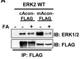
Chromatin immunoprecipitation (ChIP)
- ChIP was performed using the EZ-ChIP kit.
- The isolated calvaria cells were differentiated by treatment with 150 ng/ml rhBMP-2 for 2 days and harvested for analysis.
- Immunoprecipitations were carried out using the mouse anti-FLAG antibody and primers for analysis were designed to amplify the osteocalcin binding element; forward primer
5′-CTGAACTGGGCAAATGAGGACA-3′ , reverse primer5′-AGGGGATGCTGCCAGGACTAAT-3′ .
Micromass Mesodermal Cultures
- The effect of BMP modulators and BMP2 were analyzed by adding recombinant protein to the medium in 24 hr cultures.
- Treatments were maintained for another 24 hr period.
- After testing different protein concentrations we selected the following: human recombinant BMP2 200 ngr/ml ; human recombinant NOGGIN, 200 ngr/ml ; human recombinant CHL-1, 2400 ngr/ml ; mouse recombinant CHL-2 1200 ngr/ml ; human recombinant TSG 1000 ngr/ml ; mouse recombinant AN 3000 ngr/ml ; Follistatin 800 ngr/ml .
- After these treatments we analyzed by Q-PCR changes in the expression of cartilage markers (
Sox9 ,2 ; andBmpr1b ), fibrogenic markers (, 1 and), and joint markers ( Activinβα ,Gdf5 , and). - The selected genes are well known markers of the corresponding morpho-developmental processes.
- Only
Jaws has not been used very often as joint marker, but it has been shown that it is essential for the formation of interphalangeal joints
2.7. Multilineage Differentiation Assays
- Adipogenic, osteogenic, and chondrogenic differentiations were performed as previously reported [2 and maintained in the growth medium until confluence.
- Culture medium was then substituted with the induction one, composed by
α MEM supplemented with 10 nM dexamethasone , 10 mMβ -glycerol phosphate , 0.1 mM L-ascorbic acid-2-phosphate , 10% defined FBS , 1% L-glutamine , and 1% penicillin/streptomycin . - Induction was maintained for 14 days and, from the seventh day, 100 ng/mL BMP-2 was added.
- Confirmation of osteogenic differentiation was performed through Alizarin Red staining combined by real-time qPCR analysis (described below).
- At the end of the induction period, culture wells were washed briefly with a buffer solution containing 20 mM Tris-HCl and 150 mM NaCl in water.
- Cells were then fixed and dried with cooled absolute methanol for 2 minutes.
- After a washing step with ddH2O, cells were stained by 1.5% Alizarin Red aqueous solution pH 4.0–4.2 for 5 minutes and washed again first with ddH2O…
Chondrogenic differentiation
- Human ESCs and iPSCs were differentiated into chondrocytes using micro-mass culture.
- Briefly, ESCs and iPSCs were cultured at high-density, ~1.0 × 10⁵ cells per 10 μL × 7 spots in 6 cm culture dish for 3 hours.
- After incubation, medium was added to each dish without dissociating the cell drops.
- For differentiating pluripotent stem cells into chondrocytes, the differentiation medium contained: DMEM , 1% non-essential amino acids , 50 U/ml penicillin and 50 μg/ml streptomycin , 0.1 mM 2-mercaptoethanol , 1% ITS , 1% FBS , 10 ng/ml TGF-β1 , 10 ng/ml BMP-2 , and 50 μg/ml Ascorbic acid .
- The medium was changed every 2 days.
- After 5 days, aggregates were physically separated and transferred to suspension culture in 6 cm petri dishes containing differentiation media 1% ITS, 1% FBS, 10 ng/ml BMP-2 and 50 μg/ml ascorbic acid.
Cell culture
- Mouse pre-osteoblastic cells (MC3T3-E1, from ATCC) were cultured in Alpha-MEM medium supplemented with 10% fetal calf serum (FCS) and 1% penicillin/streptomycin.
- All cells were used at a low passage number (passage 4), and subconfluent cultures were used; the cells were plated at 10,000 cells/cm2 for experiments.
- For the soluble BMP-2 protein induction mode, the osteoblast precursors were cultured on native polymer substrates and were exposed to 300 ng/mL of recombinant human BMP-2 -containing media for 24 hours.
- The synthetic siRNA oligonucleotides specific for regions in the Runx2 mRNA were designed and synthesized by Santa Cruz Biotechnology (sc-37146).
- The siRNA oligonucleotide was screened for its silencing effect and was initially tested for its ability to knockdown the expression of Runx2 using RT-PCR and immunoblotting.
- The pharmacological agents used were 1 µM cytochalasin D and 50 nM jasplakinolide .
- The osteoblast precursors were exposed to each pharmacological agent for 1 hour, 4 h after seeding on a modified polymer.
- The cell proliferation…
Differentiation assays
-
Cultured ASC were tested for their ability to differentiate into the main mesodermal lineages as previously described [
l -ascorbic acid-2-phosphate (0.1 mM), beta-glycerol phosphate (2 mM) (all) and bone morphogenic protein BMP2 (100 ng/ml, , , ). - After 2 weeks of induction, differentiated ASC and controls were stained with ddH2o 0.5 % Alizarin Red-S (v/v).
- ASC were induced towards the adipogenic lineage using DMEM with addition of 1 % P/S, 10 % rabbit serum and 5 % horse serum supplemented with dexamethasone (1 mM), indomethacin (60 mM), rh-insulin (10 mM) and isobutylmethylxantine (0,5 mM; all from).
- ASC were maintained in differentiation media for 10 days and visualization of adipocyte differentiation was performed with Oil Red O .
Induced pluripotent stem cell-directed differentiation
- For hepatocyte differentiation, post-excised iPSCs were grown on Matrigel as stated above until reaching a 60 to 70% confluence upon which endoderm induction was initiated by replacing the post-excised iPSCs for 24 hours with RPMI 1640 medium , supplemented with 0.5 mg/ml albumin fraction V , and 100 ng/ml Activin A .
- On the following 2 days, 0.1 and 1% insulin–transferrin–selenium were added to the medium, respectively.
- Post-excised iPSCs were then cultured in hepatocyte culture medium containing 30 ng/ml fibroblast growth factor-4 and 20 ng/ml BMP2 for 4 days.
- The now-differentiated cells were then incubated in hepatocyte culture medium'>medium containing 20 ng/ml hematopoietic growth factor and 20 ng/ml keratinocyte growth factor for 6 days, in hepatocyte culture medium'>medium containing 10 ng/ml oncostatin-M plus 0.1 μM dexamethasone…