Goat Anti Mouse IGF-I polyclonal, antigen affinity
Category: Antigen Affinity Purified$150.00 – $190.00
Description
Accession
P05017
Applications
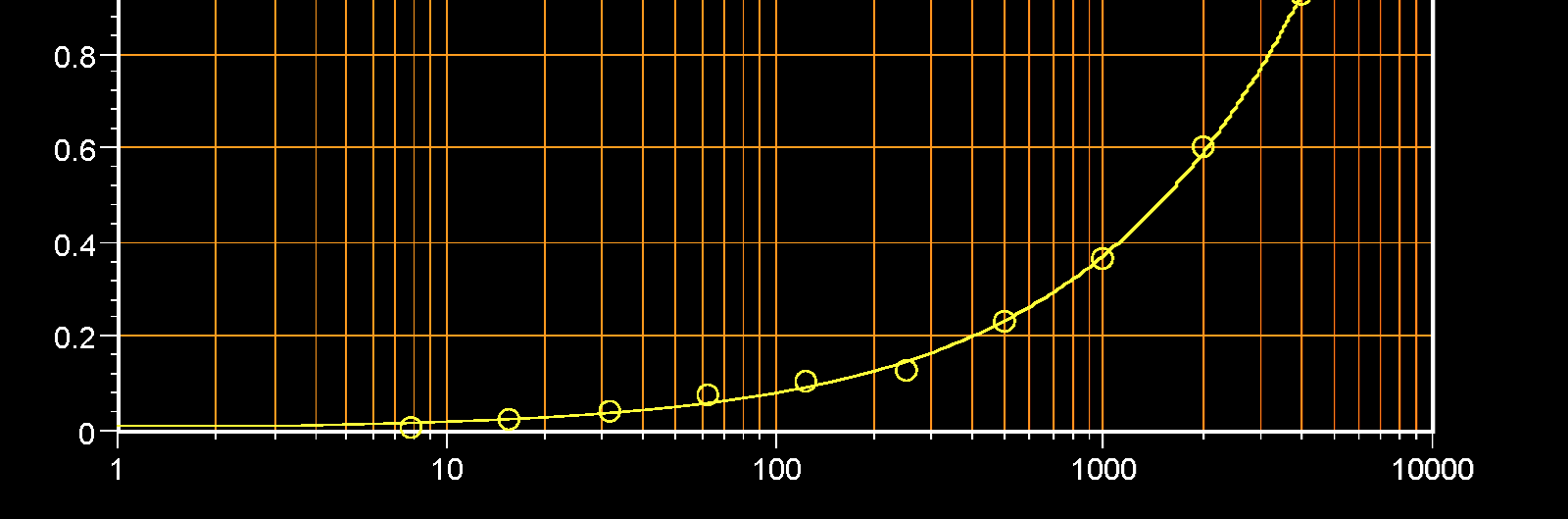
ELISA:This antibody can be used at 2 μg/mL jointly with biotinylated Mouse IGF-I antibody to detect Mouse IGF-I. The detection limit for recombinant Mouse IGF-I is approximately 0.2 ng/well.
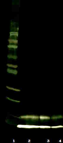
Western Blot:This antibody can be used at 0.2 μg/mL with the appropriate secondary reagents to detect Mouse IGF-I. The detection limit for recombinant Mouse IGF-I is approximately 0.2 ng/lane.
Source
This antibody was produced in Goat immunized with recombinant Mouse IGF-I.
Species reactivity
Mouse
Purification
The specific antibody was purified from Goat sera by using immobilized recombinant Mouse IGF-I affinity chromatography.
Presentation
Lyophilized from PBS, pH 7.2.
Storage
The lyophilized antibody is stable for at least 1 year from date of receipt at -20° C. Upon reconstitution, this antibody can be stored in working aliquots at 2° - 8° C for one month, or at -20° C for 12 months without detectable loss of activity. Avoid repeated freeze/thaw cycles.
Usage
This antibody product is for research purposes only.It may not be used for therapeutics or diagnostic purposes.
Molecular function
Methods
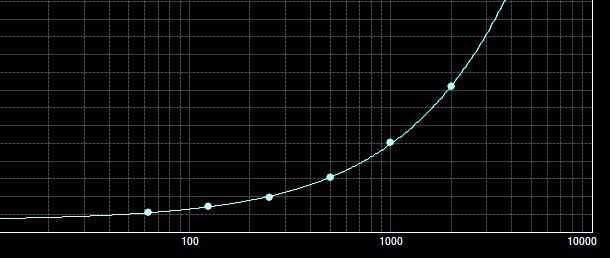
Sandwich ELISA
- 96-well maxisorp plates were coated with the capture antibody overnight, then blocked with 5% FCS in PBS for 1 hour, then washed.
- Then, the protein standard was added, and plates were incubated for 2 hrs at 37°C.
- Plates were washed and incubated with a biotinylated detection antibody for 1 hour at 37°C.
- After washing, plates were incubated with HRP-conjugated streptavidin for 30 min at room temperature and washed again.
- Plates were developed using the tetramethylbenzidine peroxidase substrate system and absorbance was measured at 450-615 nm using an automated plate reader.
Western Blot
- The Recombinant immunogen was migrated by 12% SDS-PAGE and transferred to a 0.2 um PVDF membrane.
- After blocking the membrane in blocking buffer (5% milk powder in 20 mM Tris-HCl pH 7.5, 500 mM NaCl, 0.1% (v/v) Tween 20 , the membrane was incubated with primary antibody ( at 4°C overnight.
- Peroxidase-linked secondary anti-rabbit were used to detect the bound primary antibodies.
- Enhanced chemiluminescence (ECL) reagents were used to visualize